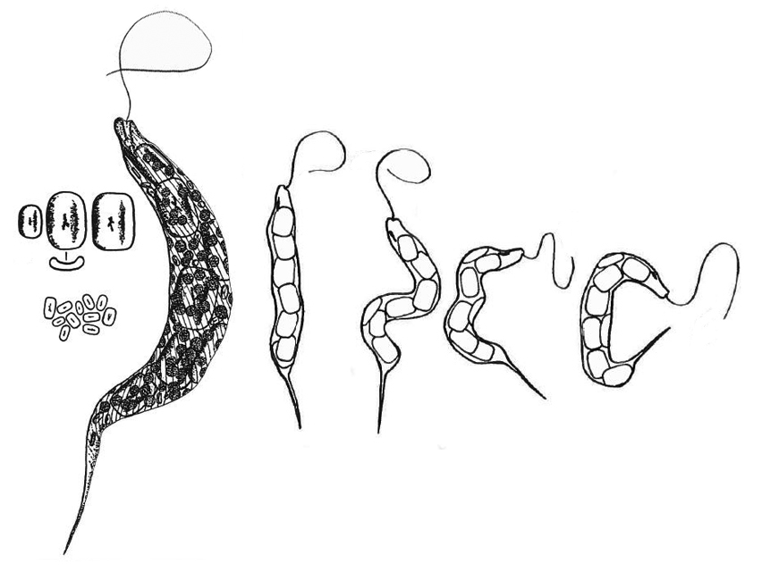
body with parallel sides, posterior end narrows to a sharp point
body often S- or C-shaped
length 120–145 µm, width 10–12 µm
pellicle with a clear striation
numerous disc-shaped chromatophores, 3–5 µm across
no pyrenoids
seven to nine prominent trough-shaped paramylon bodies with rounded corners, arranged along the longitudinal cell axis
numerous oval paramylon bodies 5–7 µm in size distributed between the prominent trough-shaped bodies
flagellum about one-sixth of body length
eyespot oval or oblong, consisting of red granules
Lepocinclis convoluta
Lepocinclis convoluta was a very rare species in Simmelried until about the year 2010, and I found it only intermittently and sporadically. After the year 2010, the population increased continuously and was at times the dominant Euglenophyceae with hundreds of specimens per milliliter. I do not know the reasons for this increase, but it was accompanied by a drastic decrease in aquatic insects and insect larvae in the Simmelried.
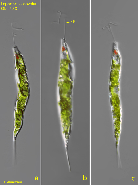
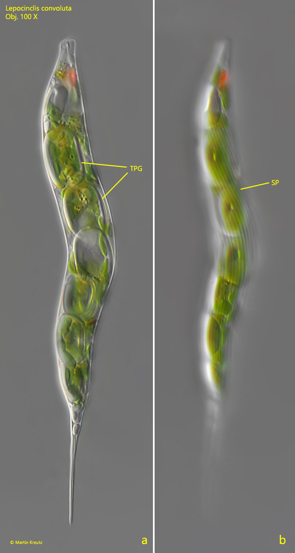
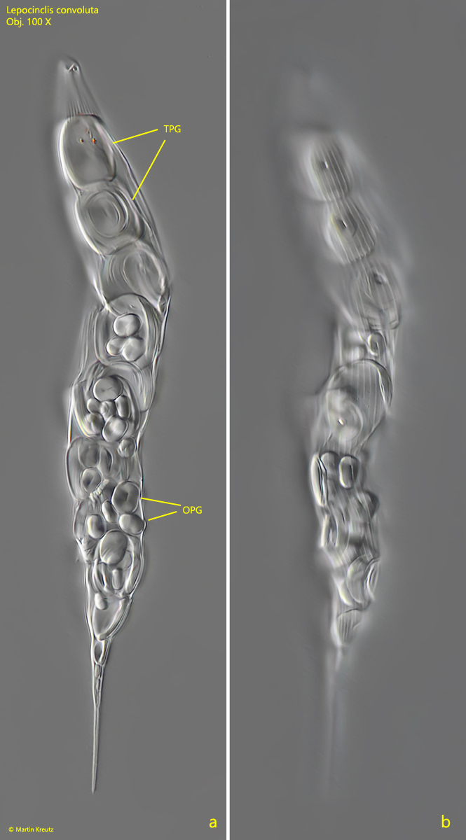
Lepocinclis convoluta is a very easy to identify because it has very large, rectangularly shaped paramylon grains with rounded corners (s. figs. 2 a-b and 3 a-b) arranged in a row in the longitudinal axis. Between them, smaller, oval-shaped paramylon grains are scattered. The chromatophores are numerous and disc-shaped (s. fig. 4). Although I had a lot of specimens available, I found freely swimming specimens only on extremely rare occasions (s. fig. 1 a-c). On these specimens I could see that the flagellum forms a lasso-shaped loop when swimming. This makes it look shorter than it is. Only in the flashed microphotos this loop can be seen (s. fig. 1 a-c). Possibly this is the reason why in the literature the flagellum is given with only one-sixth of the body length. According to my observations the flagellum is at least half as long as the body.
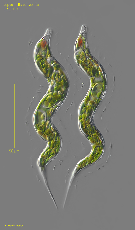
Fig. 1 a-c:Lepocinclis convoluta. L = 124- 140 µm. Three different, freely swimming specimens. Note that the flagella (F) of alle three specimens form a lasso-like end. Obj. 40 X.
Fig. 2 a-b:Lepocinclis convoluta. L = 132 µm. Two focal planes of a slightly squashed specimen. Note the prominent, trough-shaped paramylon grains (TPG) arranged in a row along the longitudinal axis and the distinct striation of the pellicle (SP). Obj. 100 X.
Fig. 3 a-b:Lepocinclis convoluta. L = 148 µm. A dead specimen in which the cytoplasm and chromatophores have decomposed, so that the trough-shaped paramylon grains (TPG), the smaller oval paramylon grains (OPG) and the striation of the pellicle are clearly visible. Obj. 100 X.
Fig. 4:Lepocinclis convoluta. Focal plane on the disc-shaped chromatophores (Chr) in a S-shaped specimen. Obj. 100 X.
Fig. 5:Lepocinclis convoluta. Two specimens shortly after cell division. During cell division the specimens are covered with a mucilaginous coat. Obj. 60 X.
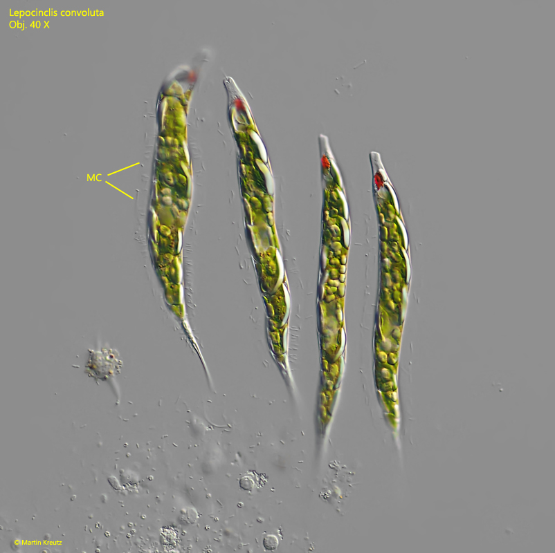
Fig. 6:Lepocinclis convoluta. Four specimens are arranged in parallel after cell division. Note the mucilaginous coat (MC) around each specimen visible by attached bacteria. Obj. 40 X.
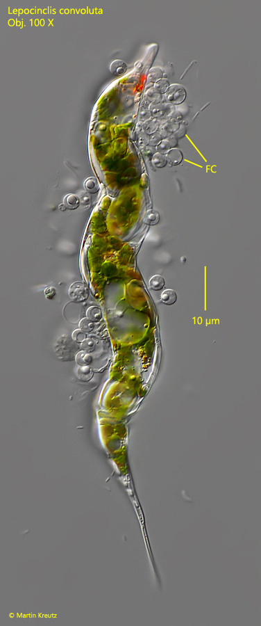
In my population of Lepocinclis convoluta I could find many cases of infestations with parasitic fungi. The infested specimens are much lighter in color, probably caused by the degradation of the chromatophores and of the paramylon due to the infestation. In these specimens I repeatedly found a granular mass (s. figs. 7 a and 8). This is likely the nucleus strongly enlarged in response to the infestation. However, it may also be the fungal cell. In one case I have found specimens covered with fungal cells (sporangia?) on the pellicle (s. fig. 9). It is unclear to me whether this was another stage of infestation with the same parasitic fungus (as shown in figs. 7a–b and 8) or another type of parasitic infestation.
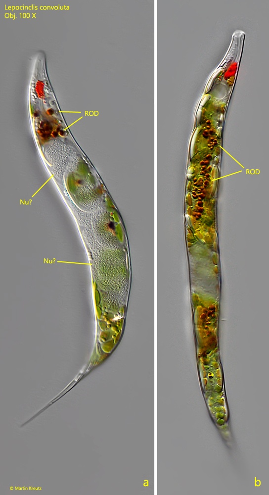
Fig. 7 a-b:Lepocinclis convoluta. Two different specimens infested by a parasitic fungus. The granulated mass is likely the nucleus (a, Nu?) enlarged in response to the infestation. In both specimens a typical accumulation of reddish oil droplets (ROD) is visible. Obj. 100 X.
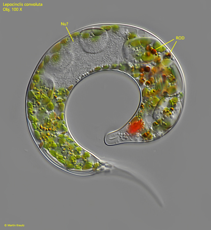
Fig. 8:Lepocinclis convoluta. A further spcimen infested by a parasitic fungus. Note that the size of the prominent trough-shaped paramylon grains is significantly decreased. NU? = likely the enlarged nucleus, ROD = reddish oil droplets. Obj. 100 X.
Fig. 9:Lepocinclis convoluta. This specimen is also infested by a parasitic fungus and covered with fungus cells (FC). It is not clear if this species of parasitic fungus is identical to the fungus which caused the infestations shown in figs. 7 a-b and 8. Obj. 100 X.