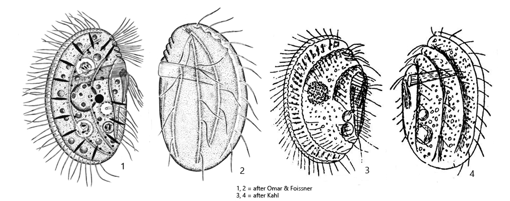
body ellipsoidal or broadly ellipsoidal, laterally flattened
ventral side is more flattened than the convex dorsal side
length 20–50 µm, width 20–35 µm
oral basket in anterior third shifted to the left side
macronucleus globular or broadly ellipsoidal, with spherical nucleoli
micronucleus globular, located at ventral side of macronucleus
extrusomes fusiform, 3–5 µm long
each 4 kineties on left and right side
cilia on left side widely spaced
contractile vacuole shortly below cell equator, near oral basket
short tube between contractile vacuole and ventral side
Leptopharynx costatus
Leptopharynx costatus is one of the most common ciliates in moss samples. If moss samples are doused with rainwater, Leptopharynx costatus can be detected with high probability after a few days.
At first glance, the ciliate resembles Microthorax, but Microthorax has the mouth opening at the posterior end and no oral basket. The body shape is quite variable. Broader and more elongated specimens occur together in the same sample. The specimens in my population were between 25–38 µm long.
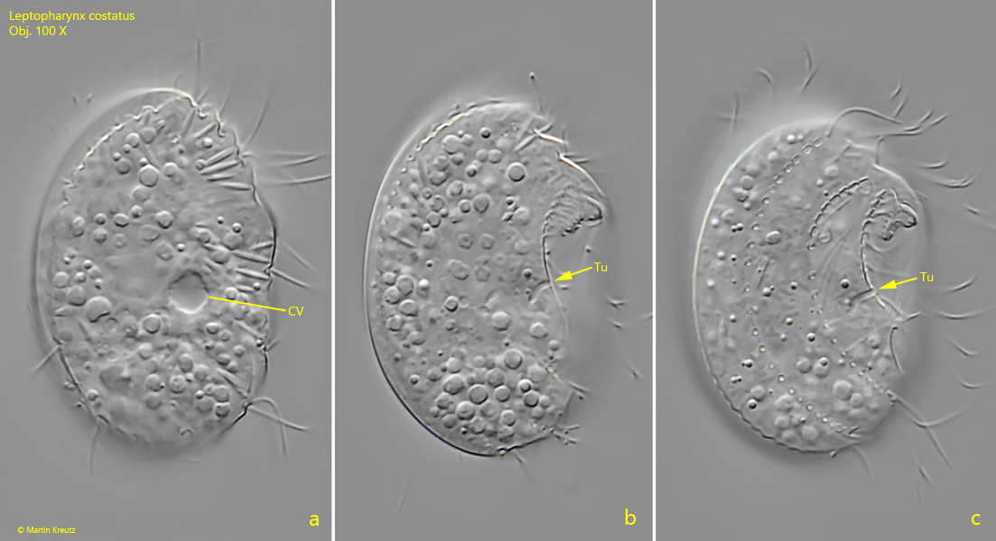
There is a thin tube between the contractile vacuole and the ventral side, which was already described by Kahl (1935). It serves to empty the contractile vacuole and opens on the ventral side below the oral basket. This fine tube is not easy to find, which once again shows how precisely Kahl observed. By Omar & Foissner (2012), this tube was only visible and drawn after silver impregnation (s. drawing 1, above). I was able to recognize the tube in a living specimen (s. fig. 3 a-c). It is bent to the right side and opens approximately in the middle of the body on the ventral side.
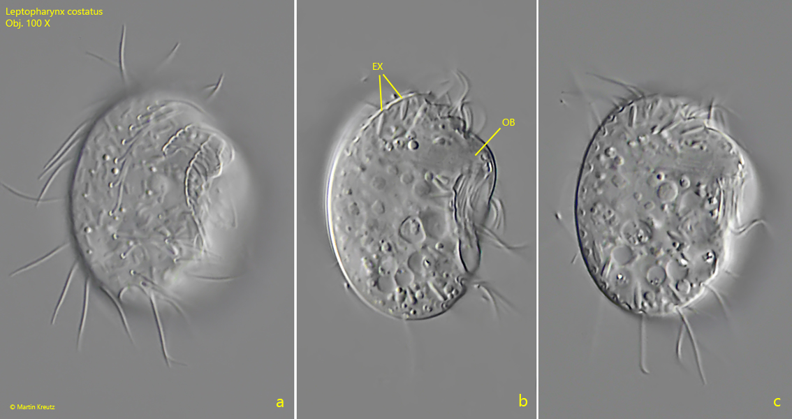
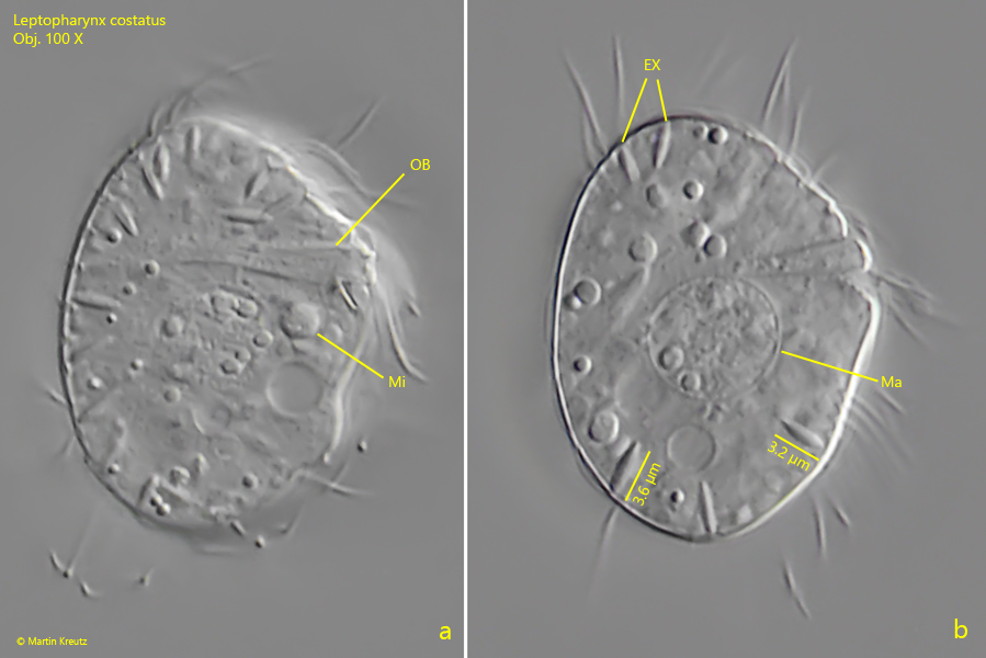
Fig. 1 a-c:Leptopharynx costatus. L = 27 µm. Three focal planes of a freely swimming specimen from right. EX = extrusomes, OB = oral basket. Obj. 100 X.
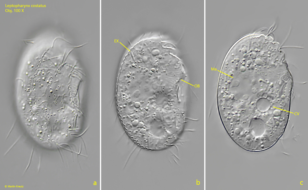
Fig. 2 a-c:Leptopharynx costatus. L = 36 µm. A slightly squashed specimen from right. Note the delicate rods of the oral basket (OB). CV = contractile vacuole, EX = extrusomes, Ma = macronucleus. Obj. 100 X.
Fig. 3 a-c:Leptopharynx costatus. L = 38 µm. Three focal planes from right for visualization of the short tube (Tu) between the contractile vacuole and the ventral side. The tube is slightly curved to the right side and open to the ventral side below the oral basket. Obj. 100 X.
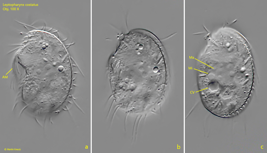
Fig. 4 a-c:Leptopharynx costatus. L = 29 µm. A freely swimming specimen from left. AM = elongated cilia of the adoral membranelles, CV = contractile vacuole, Ma = macronucleus, Mi = micronucleus. Obj. 100 X.
Fig. 5 a-b: Leptopharynx costatus. Two focal planes of a strongly squashed specimen. Note the fusiform extrusomes (EX) with a lengths of 3.2–3.8 µm. Ma = macronucleus, Mi = micronucleus, OB = oral basket. Obj. 100 X.
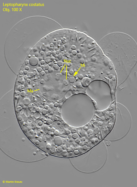
Fig. 6: Leptopharynx costatus. A second strongly squashed specimen. Ma = macronucleus, Mi = micronucleus, Nuc = spherical nucleoli. Obj. 100 X.
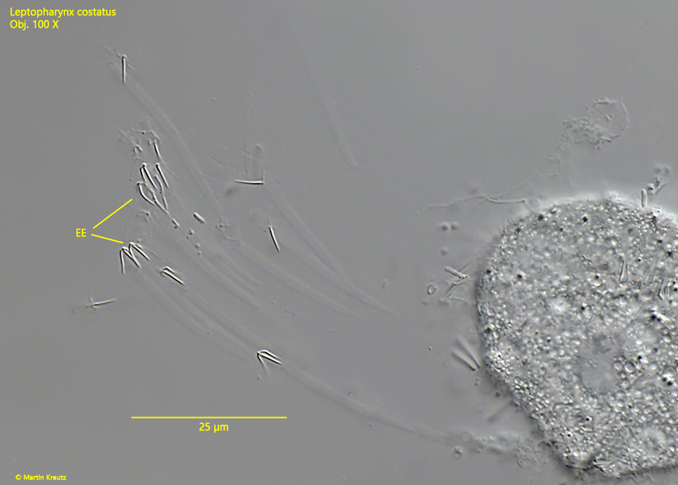
Fig. 7: Leptopharynx costatus. The ejected extrusomes (EE) are 30–40 µm long with a gelatinous stalk and a head of 4 rod-shaped arms. Obj. 100 X.