girdle of 16–22 µm long extrusomes located in mid-body region
posterior half of body covered with polygonal plates
CV subterminal (sometimes not present)
tubular neoformation organelle in posterior half
colored green by sequestered chloroplasts (functional chloroplasts from ingested algae)
macronucleus globular or ellipsoidal with 1–2 micronuclei
fast swimmer, stops sometimes and rotates around longitudinal axis
Limnostrombidium viride
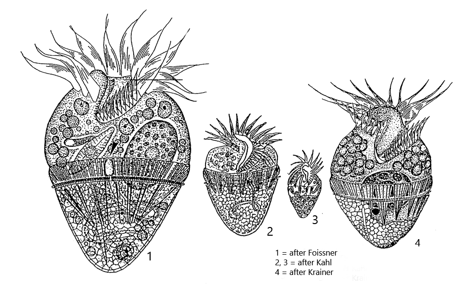
Although Limnostrombidium viride is a planktonic ciliate, I regularly find it even in sites that are not actually classic pelagic waters. However, the specimens are often found in or around floating plants. In samples the specimen then collect at the surface. Limnostrombidium viride can be easily identified by the green or yellow-green color. It is not caused by symbiotic algae, as in many other ciliates, but by their chloroplasts. That is, the phagocytosed algae are digested except for the chloroplasts, which remain fully functional and are then transported to the cell surface. By these “stolen” chloroplasts (s. fig. 3) Limnostrombidium viride can also be distinguished from the similar species Limnostrombidium pelagica, which does not possess these chloroplasts. In addition, Limnostrombidium viride has some other interesting features. For example, the posterior part of the body is covered by a thin sheath of colorless polygonal plates, which are difficult to detect in the unsquashed specimen (s. figs. 5 and 6). In addition, a tubular organelle is present in the posterior half of the body, which is called the “neoformation organelle” (s. fig. 3). It is thought to serve osmoregulation. This has been concluded because in many specimens no contractile vacuole is detectable.
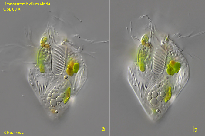
Fig. 1 a-b:Limnostrombidium viride. L = 58 µm. A free-swimming specimen in two slightly different focal planes. Obj. 60 X.
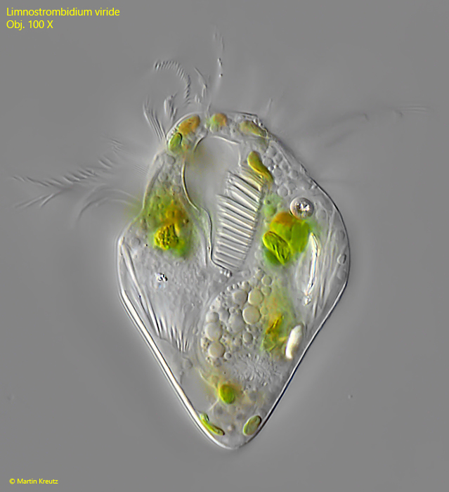
Fig. 2:Limnostrombidium viride. L = 58 µm. Ventral view of a slightly squashed specimen. Obj. 100 X.
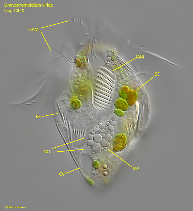
Fig. 3:Limnostrombidium viride. L = 58 µm. Ventral view of a slightly squashed specimen. CV = contractile vacuole, EX = extrusomes, IAM = inner adoral membranelles, MA = macronucleus, NO = neoformation organelle, OAM = outer adoral membranelles. Obj. 100 X.
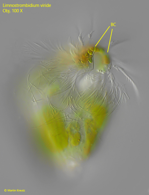
Fig. 4:Limnostrombidium viride. L = 58 µm. Ventral view of the almost circular buccal cavity (BC). Obj. 100 X.
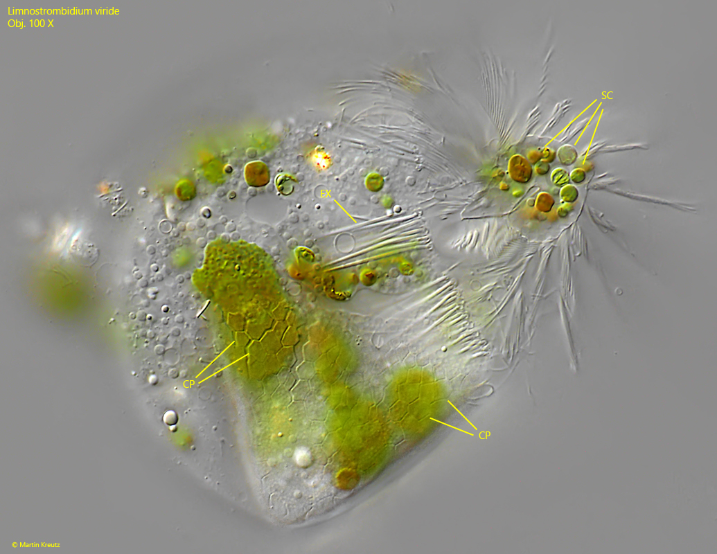
Fig. 5:Limnostrombidium viride. A squashed specimen with focal plane on the polygonal cortical plates (CP) covering the posterior half of the body. In the buccal cavity some of the sequestered chloroplasts (SC) are visible. Obj. 100 X.
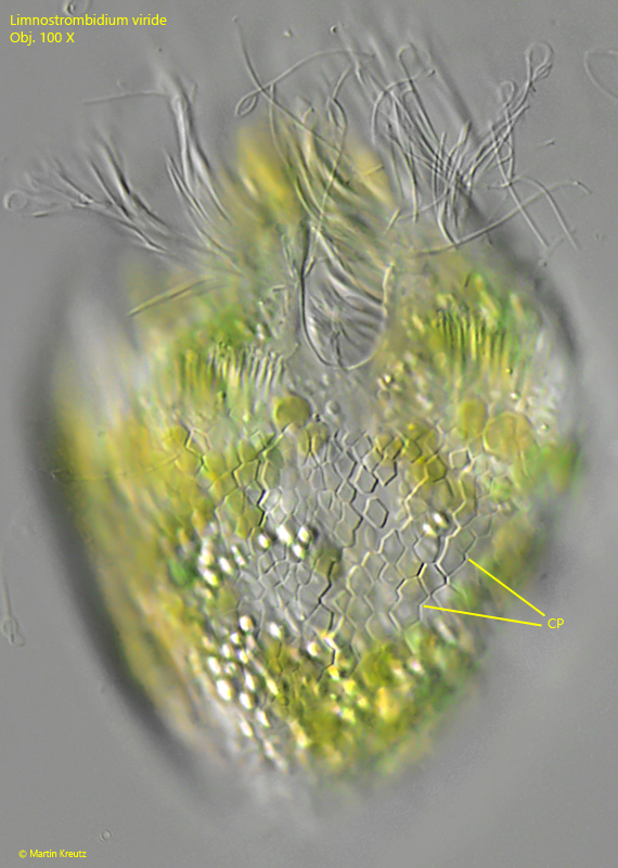
Fig. 6:Limnostrombidium viride. A second squashed specimen with focal plane on the polygonal cortical plates (CP). Obj. 100 X.
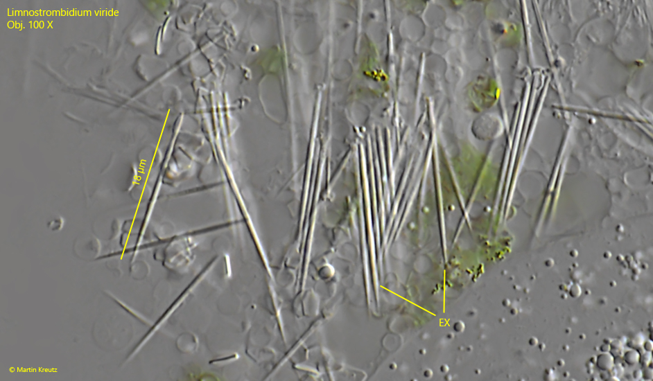
Fig. 7:Limnostrombidium viride. The extrusomes (EX) are 18 µm long with a tapered and pointed end. Obj. 100 X.