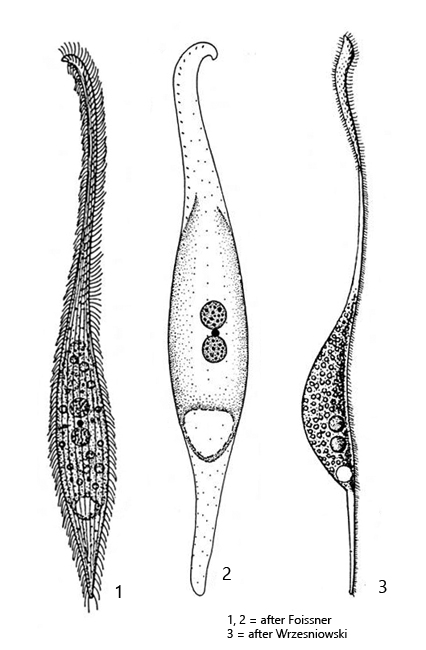
body slenderly lanceolate and flattened, ventral side convex, dorsal side straight
posterior end norrowly rounded or tail-like
neck stretched more than body length, bent dorsally
length 200–300 µm
left side with some rows of short bristles
right side with 6–13 rows of cilia
dorsal brush of club-shaped, short cilia
two spherical macronuclei
one spherical micronucleus between the macronuclei
extrusomes rod-shaped, slightly curved, 6–8 µm long
extrusomes arranged mainly along oral cleft
contractile subterminal, at the end of a left-sided bulge
sometimes a second contractile vacuole in anterior third
Litonotus cygnus
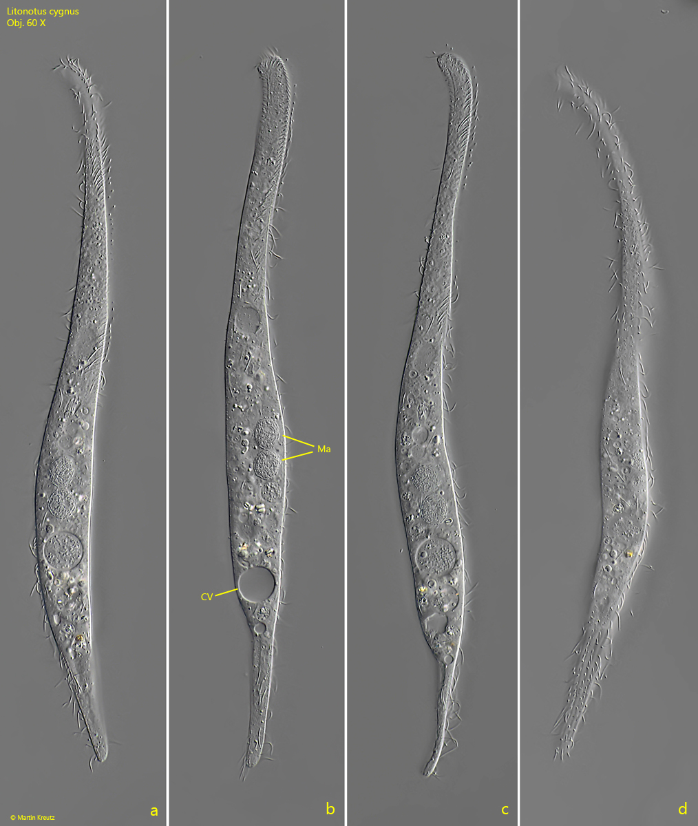
I find Litonotus cygnus in almost all of my sampling sites. The ciliate is difficult to observe in fresh specimens, but it likes to settle on the floating coverslip. Litonotus cygnus then glides along it with its right, ciliated side.
Litonotus cygnus is easy to identify. It is the largest of all Litonotus species. Typical are the two spherical macronuclei, which can be recognized even at low magnifications. The neck is very long and, especially at the front end, is clearly bent backwards and slightly widened. The posterior end is almost always tail-shaped in my population. I have only rarely found narrowly rounded forms. The extrusomes are found almost exclusively on the ventral side of the neck. This is where the oral cleft is located, but it is only visible during feeding.
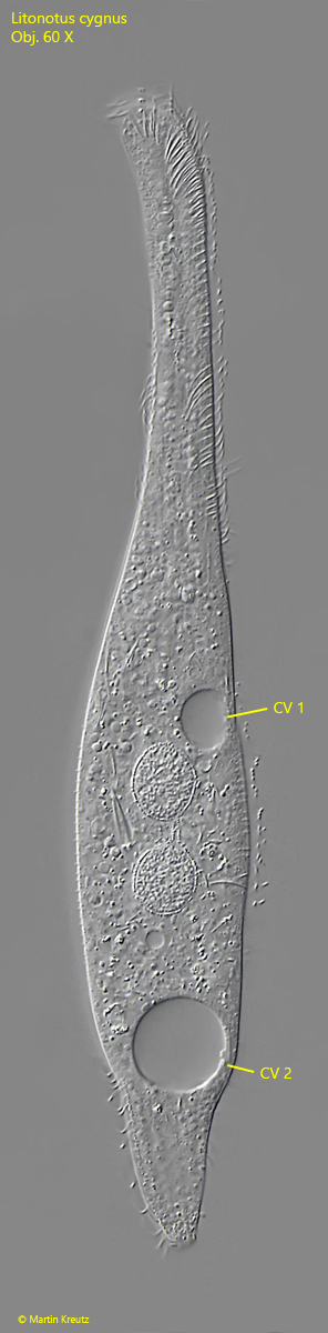
In rare cases, in addition to the subterminal contractile vacuole, a second, smaller one is visible in the anterior third (s. fig. 3). It is less active than the subterminal contractile vacuole and is particularly visible in slightly compressed specimens.
Fig. 1 a-d:Litonotus cygnus. L = 180 µm. Different focal planes of a freely swimming specimen from right. CV = contractile vacuole, Ma = two spherical parts of the macronucleus. Obj. 60 X.
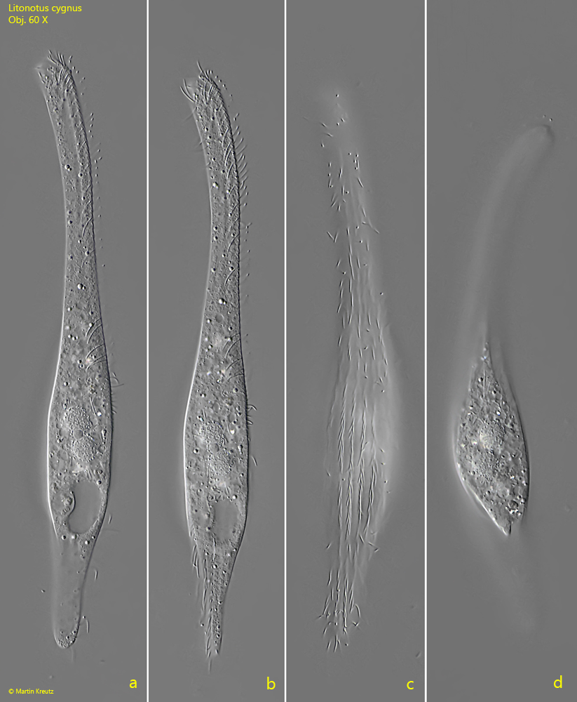
Fig. 2 a-d:Litonotus cygnus. L = 180 µm. A second specimen from right (a-c) and with focal planes on the left side (d). Obj. 60 X.
Fig. 3:Litonotus cygnus. L = 180 µm. The slightly specimen as shown in fig. 2 a-d. Note the second contractile vacuole (CV 1) in the anterior third. CV 2 = subterminal contractile vacuole. Obj. 60 X.
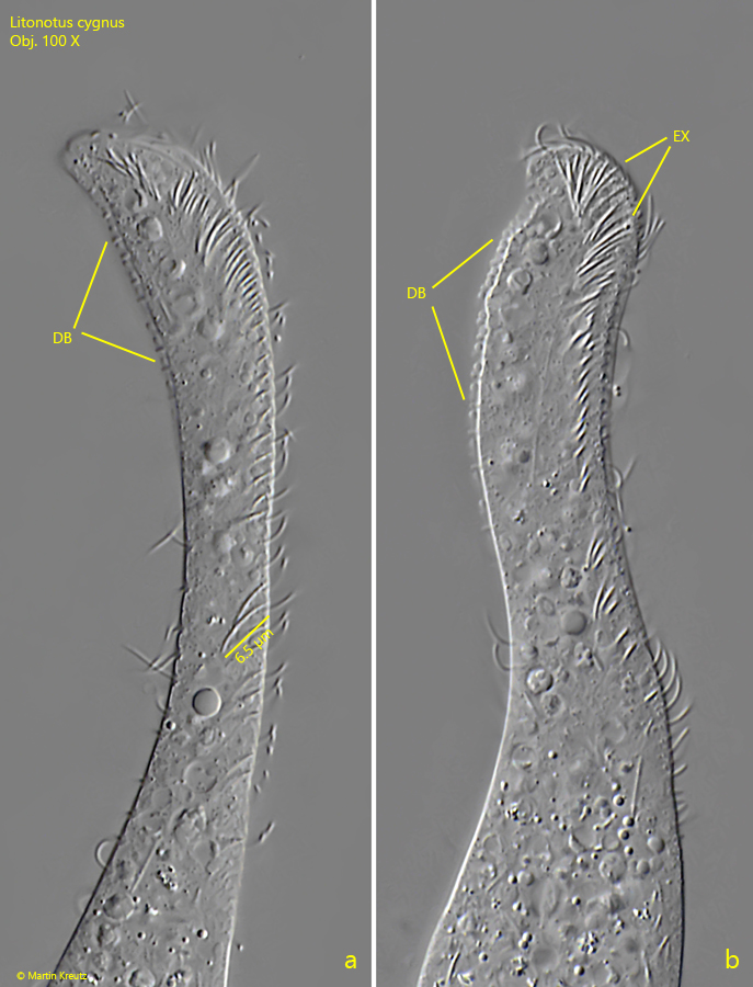
Fig. 4 a-b: Litonotus cygnus. The anterior end with the fringe of extrusomes (EX) and the dosal brush (DB) in detail. The extrusomes are curved and 6.5 µm long. Obj. 100 X.
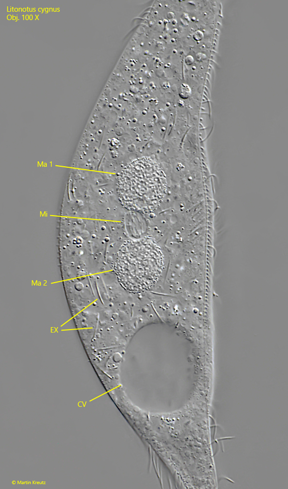
Fig. 5: Litonotus cygnus. The nuclear apparatus is consisting of two sperical macronuclei (Ma 1 , Ma 2 ) and a spherical micronucleus in between. Obj. 100 X.