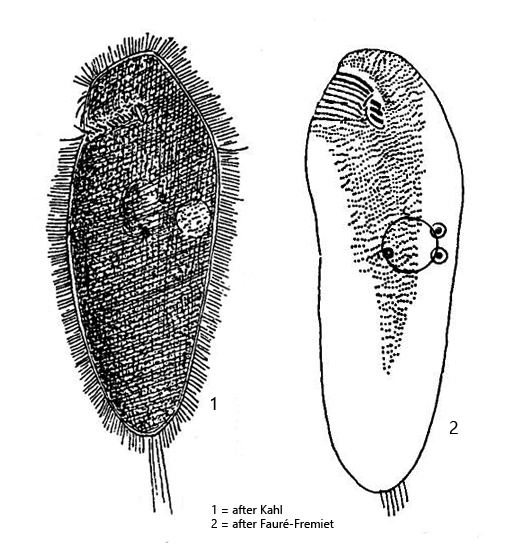
body obovate, anterior end slightly beveled and indistinctly capped
length 120–200 µm (mostly about 160 µm)
oral apparatus small, subapical, with 3 adoral membranelles
apically some transverse perizonal rows (hard to see)
globular or ellipsoid macronucleus central
4–8 micronuclei adjacent to macronucleus
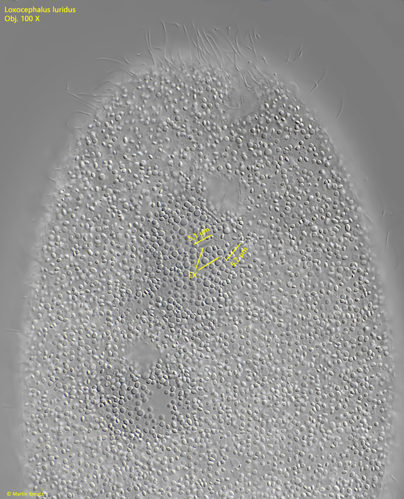
extrusomes short and spindle shaped, inconspicuous
contractile vacuole in midbody
cytoplasm filled with refractile granules, giving a dark appearance
about 120 rows of cilia
tuft of caudal cilia, about 15–20 µm long
Loxocephalus luridus
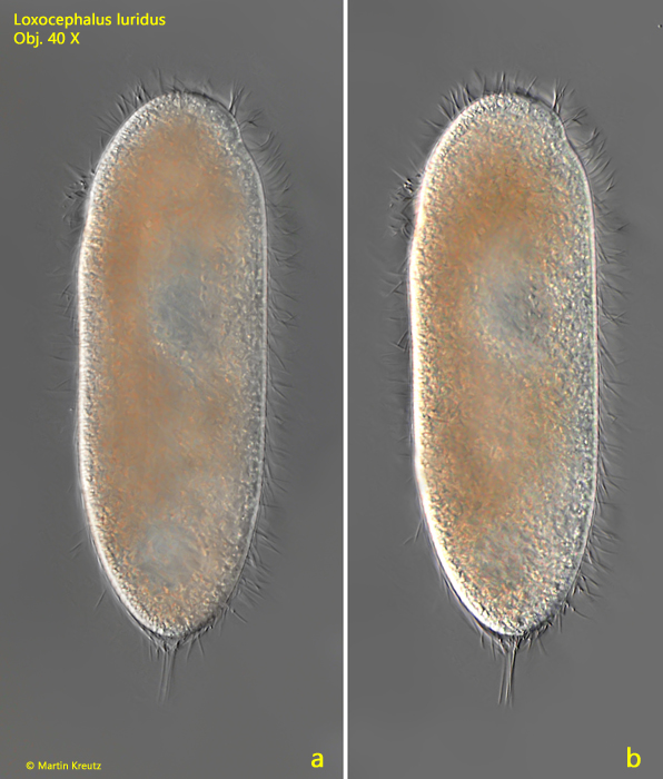
Loxocephalus luridus occurs in all my sampling sites that have a mud layer of decomposed plant material. In this layer Loxocephalus luridus is very common and associated with other anaerobic ciliates such as Metopus or Caenomorpha. At low magnifications, the specimens often appear black or dark brown due to the very dense granula in the cytoplasm. Loxocephalus luridus is a fast swimmer and is also quite sensitive when a coverslip is placed on it. It is therefore advisable to place the coverslip at a very high layer thickness and then observe the sample during the evaporation progress. The specimens often found attached to detritus flakes and can then be easily observed without cover glass pressure. The apical end of Loxocephalus luridus is slightly cap-shaped (s. fig. 1 a-b).
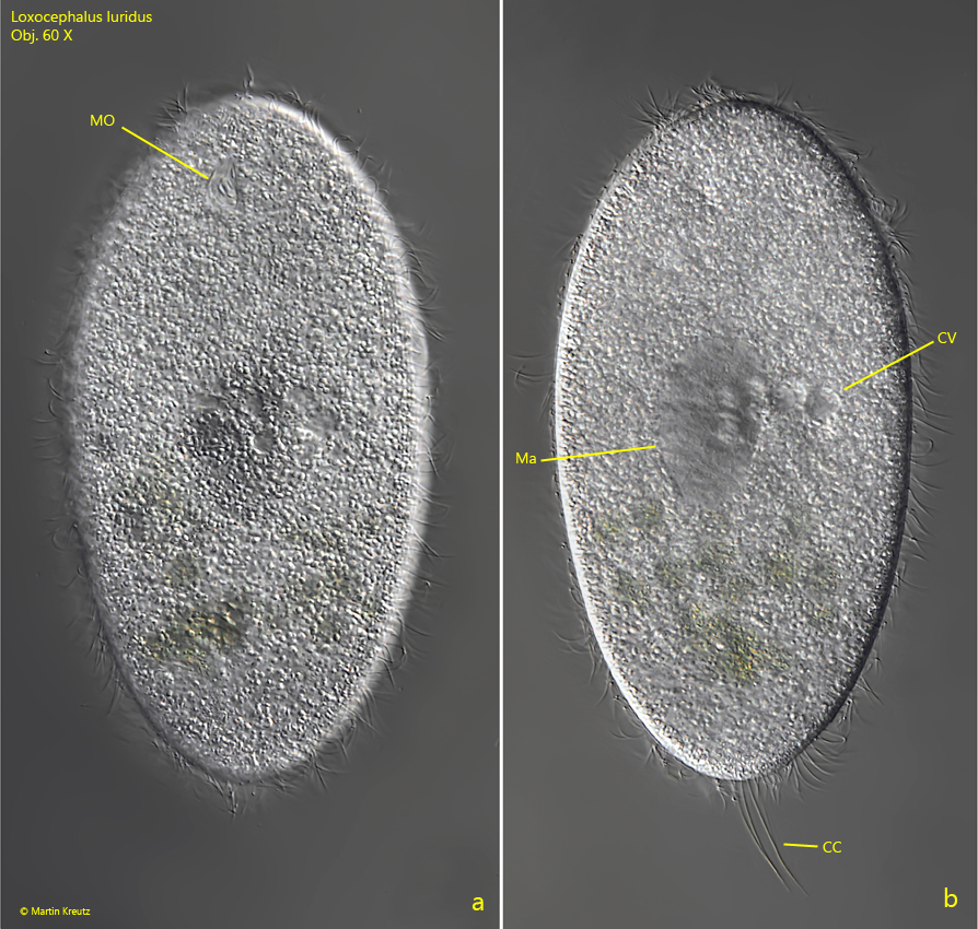
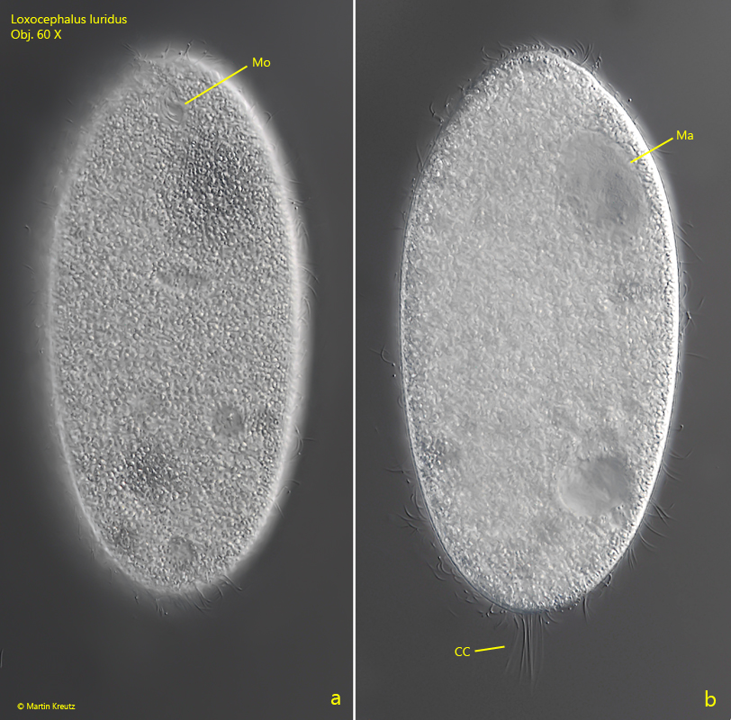
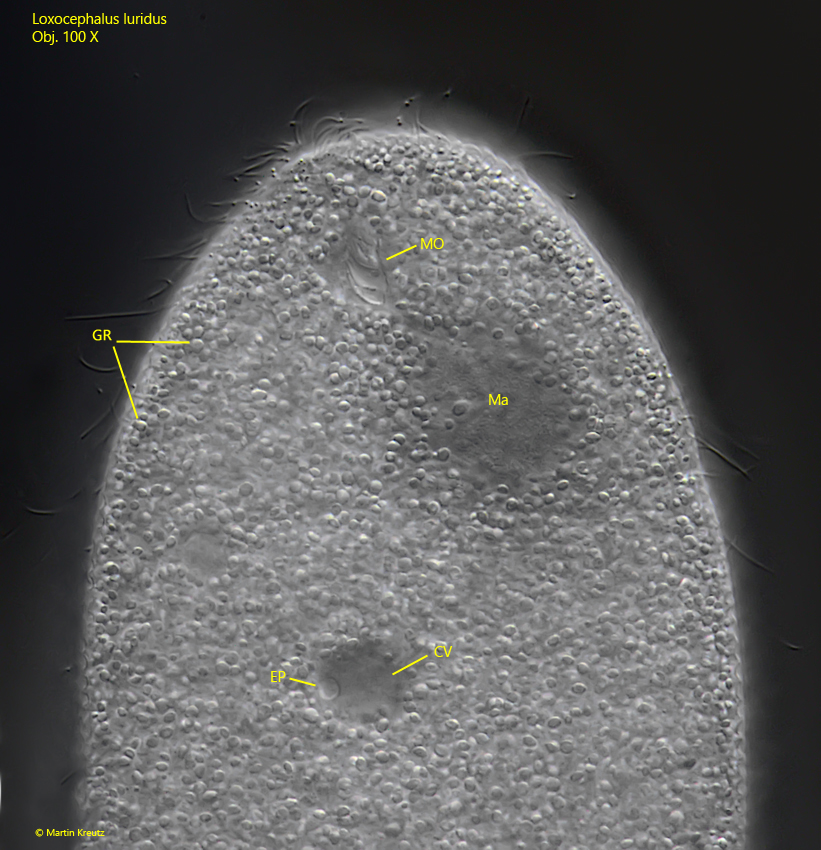
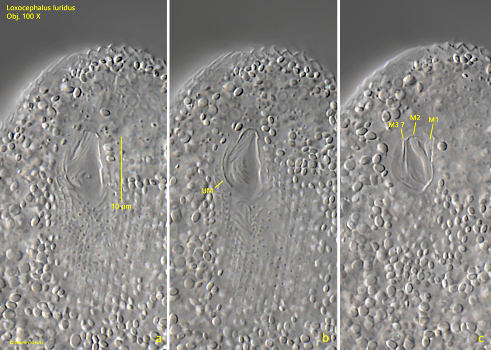
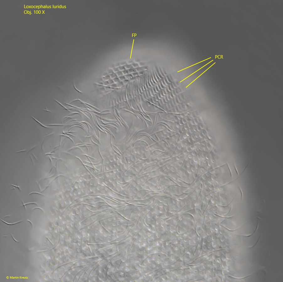
At higher magnification the granules in the cytoplasm appear irregularly shaped with a diameter of 1–3 µm (s. fig. 4). Due to the granula it difficult to recognize the inner cell structure. The round macronucleus is located in the center of the cell, but it can be easily displaced by coverslip pressure. The contractile vacuole, which has an excretory porus on the ventral side, is also located in the center of the cell (s. fig. 4). The mouth opening is extraordinarily small for a ciliate of this size. Three adoral membranelles are located one behind the other in the oral cavity, but these are difficult to recognize without silver impregnation (s. figs. 4 and 5 a-c). The preoral suture can be seen apically on the dorsal side (s. fig. 6). It runs ventrally around the right side and ends above the mouth opening. The caudal cilia are close together and lie parallel (s. figs. 2 b and 3 b). They are all about the same length. If the focal plane is on the pellicle, a honeycomb-like structure becomes visible (s. fig. 7).
Fig. 1 a-b:Loxocephalus luridus. L = 172 µm. Two focal planes of a freely swimming specimen. Note the cap-shaped anterior end. Obj. 40 X.
Fig. 2 a-b:Loxocephalus luridus. L = 162 µm. Two focal planes of a slightly squashed specimen from ventral. CC = tuft of caudal cilia, CV = contractile vacuole, Ma = macronucleus, MO = mouth opening. Obj. 60 X.
Fig. 3 a-b:Loxocephalus luridus. L = 176 µm. A second slightly squashed specimen from ventral. CC = tuft of caudal cilia, Ma = macronucleus, MO = mouth opening. Obj. 60 X.
Fig. 4:Loxocephalus luridus. The anterior half of a slightly squashed specimen from ventral. Note the excretion porus (EP) of the contractile vacuole (CV). The cytoplasm is filled with irregular shaped, refractive granules (GR) with a diameter of 1–3 µm. Ma = macronucleus, MO = mouth opening. Obj. 100 X.
Fig. 5 a-c:Loxocephalus luridus. Three focal planes of the oral apparatus. Note the adoral membranelles (M1, M2 and M3 ? = probably membranelle 3) and the undulating membrane (UM). Obj. 100 X.
Fig. 6:Loxocephalus luridus. Dorsal view of a slightly squashed specimen with the perizonal ciliary rows (PCR). Note the fielded pellicle (FP). Obj. 100 X.
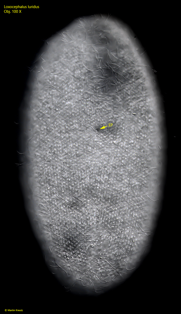
Fig. 7:Loxocephalus luridus. A slightly squashed specimen from ventral with focal plane on the fielded pellicle. EP = excretion porus. Obj. 100 X.
Fig. 8:Loxocephalus luridus. Focal plane on the granules in the cytoplasm. The granules are spherical, oval or ellipsodial but not irregularly shaped. Between the granules two spindle-shaped extrusomes (EX) are visible. Obj. 100 X.