granules arranged in rows beneath pellicle, 6–7 rows per body side
one caudal cilium
Loxocephalus moniligranulatus
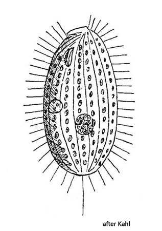
So far I have only been able to find a single specimen of Loxocephalus moniligranulatus in December 2020 in Ulmisried. The specimen came from a layer of decomposing leaves. The species is also described by Kahl as “nicht sehr verbreitet, nicht zahlreich (not very common, not numerous)” and provides only a very brief description.
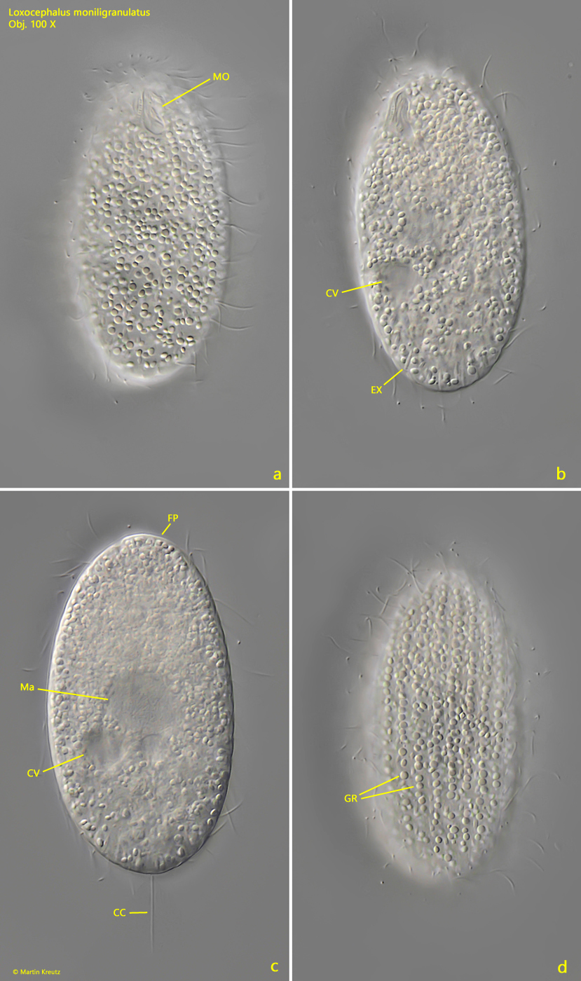
An essential characteristic of Loxocephalus moniligranulatus are the granules arranged in longitudinal rows below the pellicle (s. fig. 1 d). Inside the cell, however, the granules are disordered. According to my measurements, the granules have a diameter of 1–2 µm and appear lenticular to me. There are also scattered spindle-shaped extrusomes under the pellicle, which Kahl does not mention (s. fig. 1 b). They were 4–4.5 µm long in my specimen. The macronucleus and the contractile vacuole of Loxocephalus moniligranulatus are located near the middle of the body (s. figs. 1 b and 1 c).
The similar species Loxocephalus ellipticus also has granules arranged in longitudinal rows under the pellicle. This species is slightly smaller at around 40–50 µm and the contractile vacuole is located at the posterior end instead midbody. This makes the two species easily distinguishable.
Fig. 1 a-d:Loxocephalus moniligranulatus. L = 63 µm. Different focal planes of a slightly squashed specimen from ventral (a, b) and from dorsal (c, d). Note the refractive granules (GR) arranged in longitudinal rows beneath the pellicle (d). The globular macronucleus (Ma) and the contractile vacuole (CV) are located near midbody. CC = caudal cilium, EX = extrusomes, FP = frontal plate, MO = mouth opening. Obj. 100 X.