body elongated oval or cylindroid, often dark colored
length 50–65 µm
apical front plate present
transverse ring of long adoral cilia in anterior third
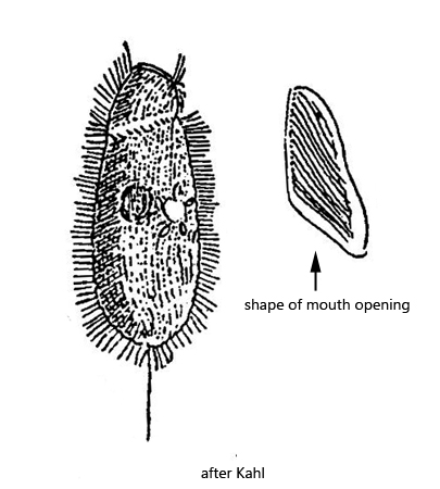
oral opening almost crescentically shaped
spherical or broadly oval macronucleus in mid-body
one contractile vacuole in mid-body
conical shaped extrusomes, 3.2–3.3 µm long
numerous granules in cytoplasm, not ring-shaped
one caudal cilium
Loxocephalus plagius
So far I have only found Loxocephalus plagius in the Simmelried. However, as it is a very common ciliate, I assume that it also occurs in my other sites with a layer of mud and rotting plant parts.
Loxocephalus plagius is very easy to confuse with Dexiotricha granulosa, as both species are about the same size. In Dexiotricha granulosa, however, the granules in the cytoplasm are ring-shaped and the cilia (inclusive the caudal cilium) are rigid and straight. In addition, the mouth opening of Dexiotricha granulosa is smaller and the left side is C-shaped. In Loxocephalus plagius the mouth opening is elongated, almost crescent-shaped and on the left edge there is a characteristic angle of about 120 °, as also drawn by Kahl (s. drawing above and figs. 1 c and 4 c).
In my population of Loxocephalus plagius I could find distinct, spindle-shaped extrusomes, which are 2.2–3.3 µm long and are arranged beneath the pellicle (s. figs. 2 a, 3 a and 5). These extrusomes are not mentioned or drawn by Kahl in his very short description.
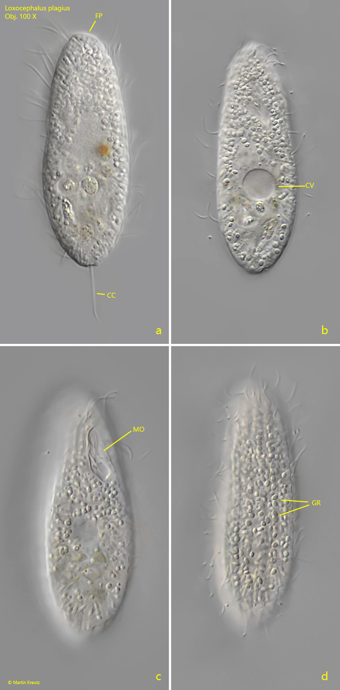
Fig. 1 a-d:Loxocephalus plagius. L = 46 µm. Different focal planes of a freely swimming specimen. Note the crecentic shape of the mouth opening (MO). CC = caudal cilium, CV = contractile vacuole, EX = extrusomes, Mi = micronucleus. Obj. 60 X.
Fig. 2 a-d:Loxocephalus plagius. L = 46 µm. Different focal planes of a second freely swimming specimen. CC = caudal cilium, EX = extrusomes, FP = frontal plate, Ma = macronucleus, Mi = micronucleus. Obj. 60 X.
Fig. 3 a-c:Loxocephalus plagius. L = 46 µm. The specimen as shown in fig. 2 a-d in detail. CC = caudal cilia, EP = excetuion porus of the contractile vacuole, EX = extrusome, Ma = macronucleus, UM = undulating membranelle. Obj. 100 X.
Fig. 4 a-d:Loxocephalus plagius. L = 53 µm. Different focal planes of a third freely swimming specimen. Note the crecentic shape of the mouth opening (MO) and the irregularly, not ring-shaped granules (GR). CC = caudal cilium, CV = contractile vacuole, FP = frontal plate. Obj. 100 X.
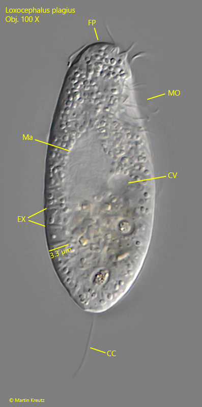
Fig. 5:Loxocephalus plagius. L = 55 µm. A slightly squashed specimen from right. CC = caudal cilium, CV = contractile vacuole, EX = extrusomes, FP = frontal plate, Ma = macronucleus, MO = mouth opening. Obj. 100 X.