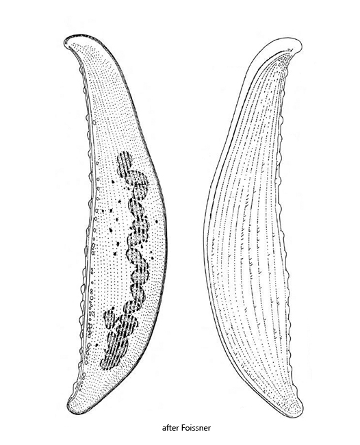
body lanceolate, knife-shaped or leaf-like, laterally flattened
length 200–700 µm (commonly 300–500 µm)
ventrally broad seam of extrusomes
dorsally warts with budles of extrusomes
right side ciliated, left side with rows of short bristles
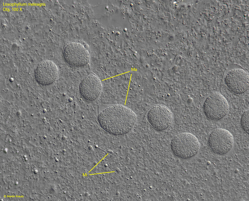
macronucleus moniliform, consisting of 16–31 nodules
7–26 cone-shaped micronuclei scattered in the plasm
contractile vacuole terminal with a long collecting canal
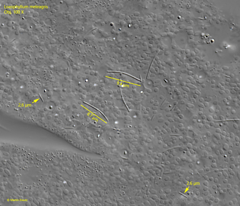
4 types of extrusomes, the largest about 12 µm long and curved
Loxophyllum meleagris
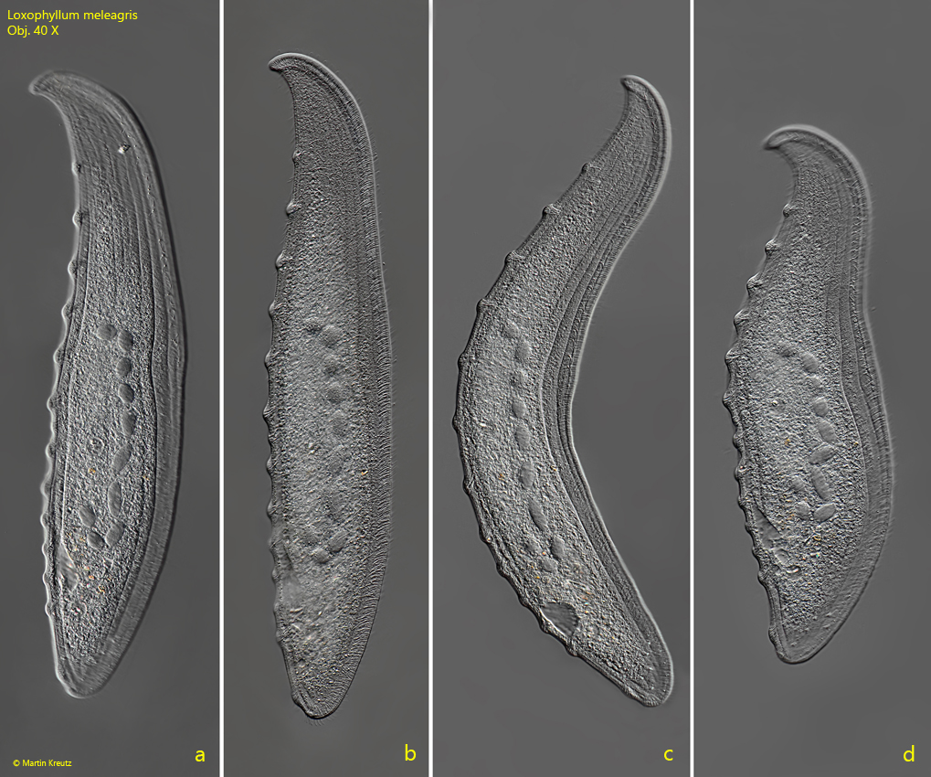
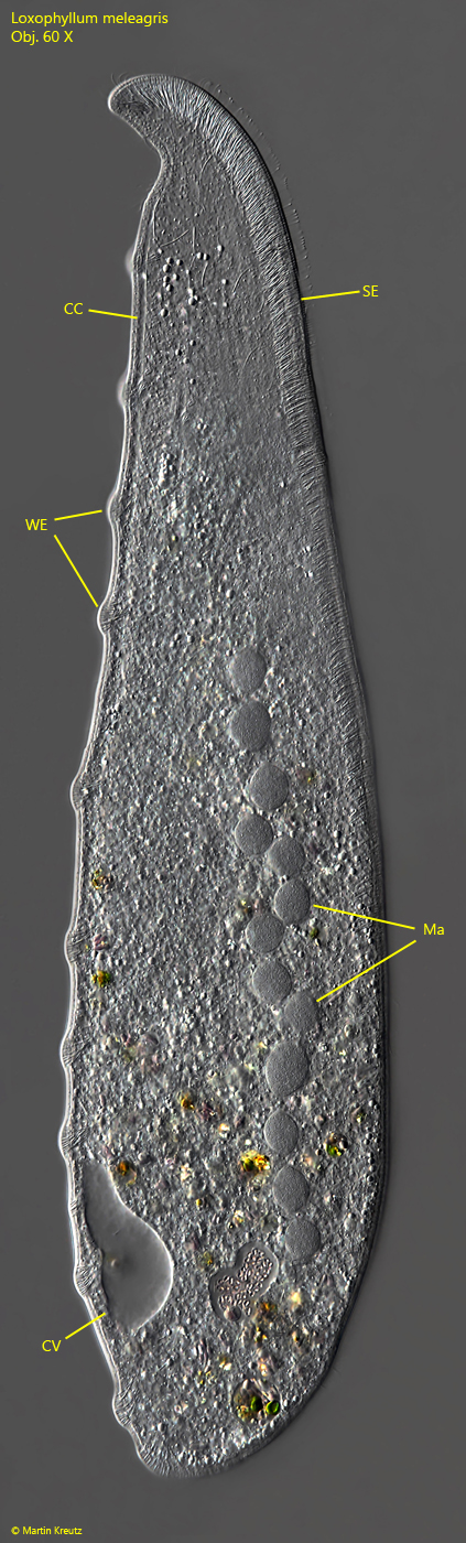
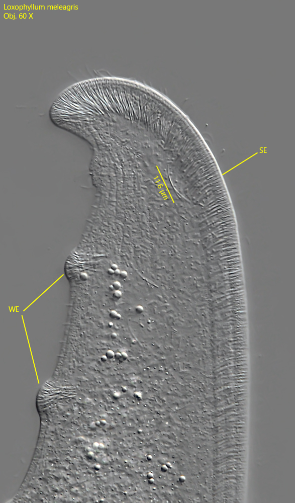
Loxophyllum meleagris is a very common ciliate that I find regularly in almost all my sites. Very often it colonizes floating coverslips, along which it glides and can be easily observed. This large ciliate feeds on rotifers and is therefore well armed with extrusomes. On its dorsal side, the warts with bundles of extrusomes typical of the genus Loxophyllum can be seen (s. fig. 2). Ventrally, densely packed extrusomes form a broad seam (s. fig. 2). The mouth opening is only visible during phagocytosis. It is located ventrally in the anterior third. The macronucleus is distinctly moniliform and easily seen, but the small, cone-shaped micronuclei are scattered in the plasma and difficult to see (s. fig. 4). The collecting duct of the terminal contractile vacuole runs along the dorsal margin (s. fig. 2). It extends almost to the apical end. Loxophyllum meleagris is continuously ciliated only on the right side. The left side appears naked by light microscopy, with the exception of rows of short bristles. Due to the strong lateral flattening Loxophyllum meleagris is very thin and therefore a photogenic species.
Fig. 1 a-d:Loxophyllum meleagris. L = 400 µm. Different stages of a gliding specimen from right. Obj. 40 X.
Fig. 2:Loxophyllum meleagris. L = 460 µm. A fully extended, knife shaped specimen from right. CC = collecting canal, CV = contractile vacuole, Ma = moniliform macronucleus, SE = ventral seam of extrusomes, WE = dorsal warts with bundles of extrusomes. Obj. 60 X.
Fig. 3:Loxophyllum meleagris. Detail of the anderior end. Note the approximately 12 µm long extrusomes, which form the ventral seam (SE) in a parallel arrangement. On the dorsal side the extrusomes are arranged in bundles to form the warts (WE). Obj. 60 X.
Fig. 4:Loxophyllum meleagris. The moniliform macronucleus (Ma) in a strongly squashed specimen. The micronuclei (Mi) are often cone-shaped and scattered in the cytoplasm. Obj. 100 X.
Fig. 5:Loxophyllum meleagris. In the cytoplasm of a strongly squashed specimen the two of the four types of extrusomes are visible. The slightly curved rods with a length of 8–10 µm and the short, straight rods with a length of 2.6 µm. Obj. 100 X.