one equatorial cirrus arises at the base of the spine
anteriorly 2 long cirri arise
adoral zone of membranelles sub-equatorial spiralling beneath the perizonal stripe
mouth opening in posterior third
macronucleus spherical or ellipsoidal with one adjacent spherical micronucleus
contractile vacuole subterminal
Ludio parvulus
Ludio parvulus is a small caenomorphid ciliate which I find frequently in the Simmelried and the Purren pond. It swims enormously fast and is coverslip-sensitive. Already Kahl mentions in his description of Ludio parvulus: “Bewegung sehr schnell…schwer festzulegen (Movement very fast……hard to immobilize under coverslip)”.
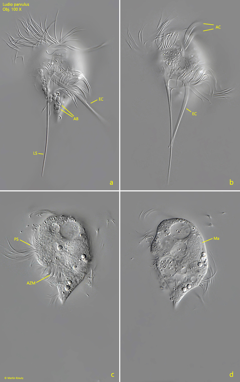
The exact structure of Ludio parvulus is difficult to observe in freely swimming specimens. One needs immobilized or chemically fixed specimens or flashed microphotographs. On the left side of the body, a long spine arises approximately in the equatorial region (s. fig. 3a), which tapers to a very fine tip. According to my observations the length of the spine can be up to 70 µm (s. fig. 2a). Also on the left side 3 cirri arise (s. fig. 4b). One cirrus in the equatorial zone arises at the base of the spine, on its right side. Two further cirri arise in the apical region of the left side (s. fig. 4 b). The perizonal stripe of cilia also originates on the left side of the body, to the left of the two apical cirri (s. fig. 4b). The perizonal stripe is running clockwise in a 360 ° turn to the posterior end, where it terminates above the mouth opening. The contractile vacuole is located subterminally, near the extended caudal tip of the body (s. fig. 3 b). The macronucleus is mostly located in the anterior third of the body (s. fig. 4 d).
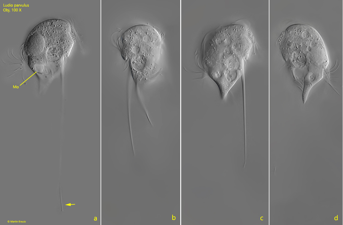
Fig. 1 a-d:Ludio parvulus. L = 32 µm. Different focal planes of a freely swimming specimen. Obj. 100 X.
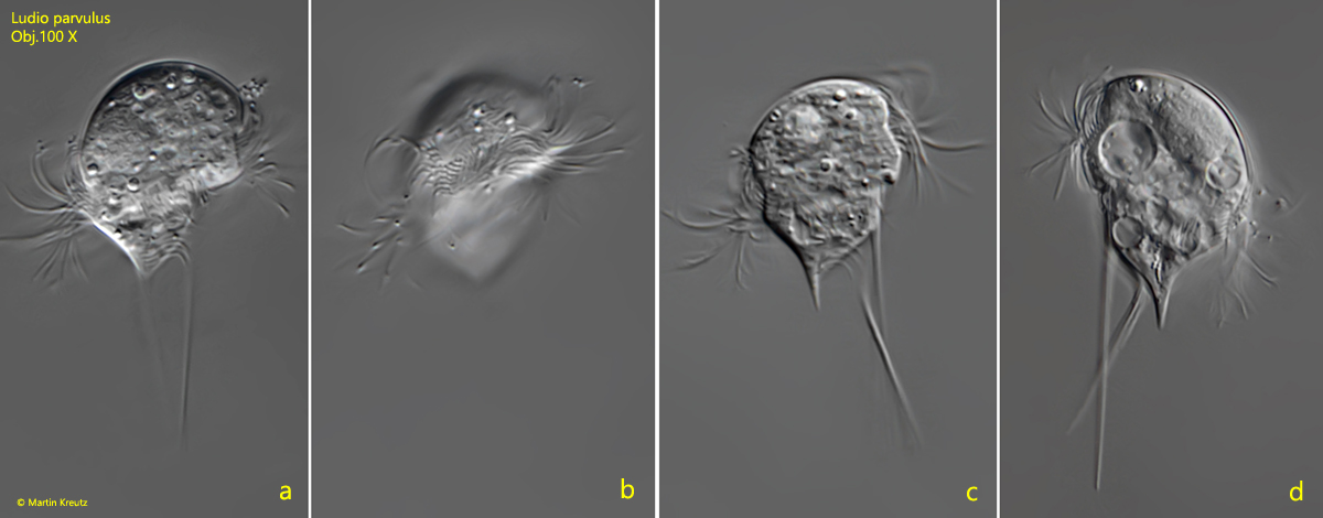
Fig. 2 a-d:Ludio parvulus. L = 37 µm. Different focal planes of a second, freely swimming specimen. Note the fine tapered end of the left spine (arrow). MO = mouth opening. Obj. 100 X.
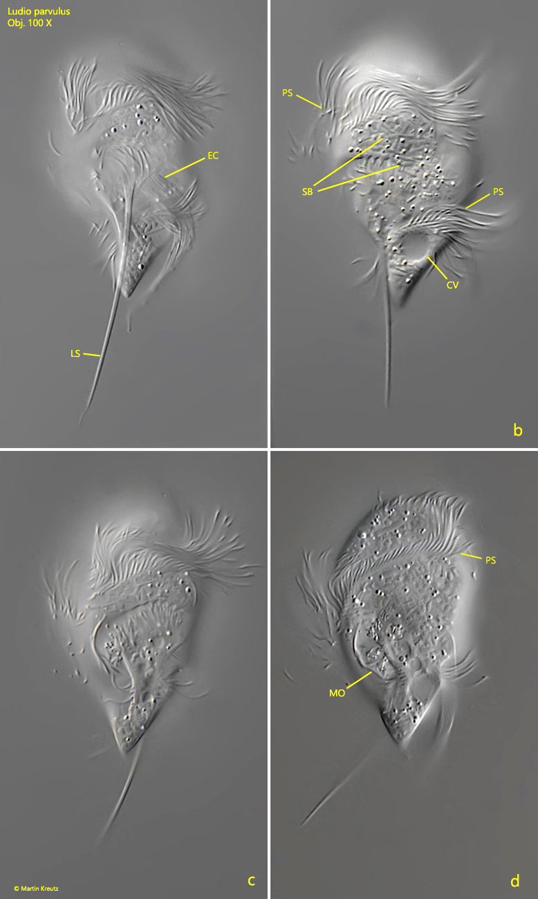
Fig. 3 a-d:Ludio parvulus. L = 40 µm. Left (a,b) and right view (c,d) of a slightly squashed specimen. CV = contractile vacuole, EC = equatorial cirrus, LS = spine at the left side, MO = mouth opening, PS = perizonal stripe of cilia, SB = symbiotic bacteria. Obj. 100 X.
Fig. 4 a-d:Ludio parvulus. L = 37 µm. Left (a, b) and ventral view (c, d) of a slightly squashed specimen. Note the arrangement of the left spine (LS), the equatorial cirrus (EC) arises at the base of the spine and the two apical cirri (AC). AB = adhering bacteria on the cell surface, AZM = adoral zone of membranelles of the mouth opening, Ma = macronucleus, PS = perizonal stripe of cilia. Obj. 100 X.
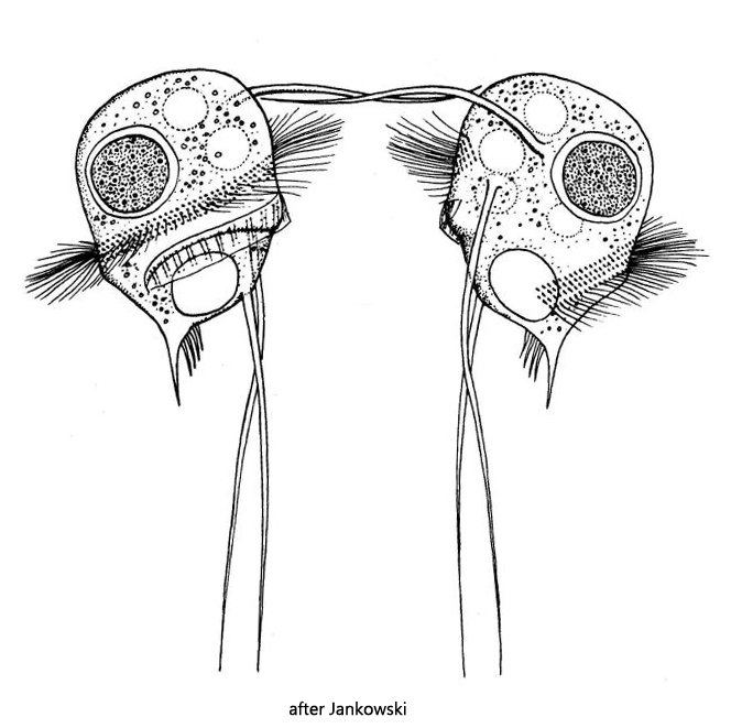
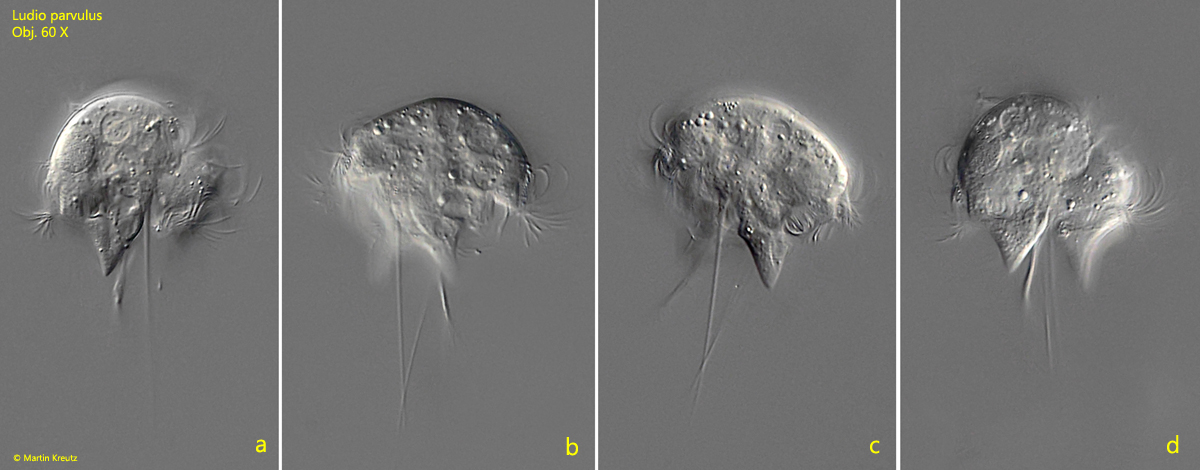
Fig. 5 a-d:Ludio parvulus. L = 35 µm. Two conjugating specimens in lateral view. Obj. 60 X.