cell spindle-shaped, apical end rounded, posterior pointed
length 23–62 µm
apical tuft of 6–8 long bristles, about 20–40 µm long
body covered with trigonal scales (hard to see)
two chloroplasts
contractile vacuoles apically located
one flagellum, about body length
cysts 6–17 µm long, oblong or broadly oval
Mallomonas akrokomos
I have only found Mallomonas akokomos once in the plankton of the Mühlhalden pond in March 2017. With a length of approx. 40 µm and a spindle-shaped body, the cells are easy to overlook. The tuft of long bristles at the apical end is characteristic. This makes the species difficult to confuse with other Mallomonas species.
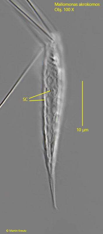
The scales are described as triangular, which should not overlap. According to my observations, they appear to be more diamond-shaped. They appear triangular due to a slight overlap (s. fig. 4). I could make out the contractile vacuoles in the middle of the body and not at the front end.
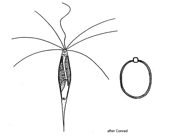
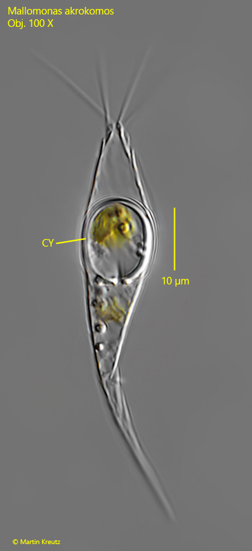
I was able to clearly identify the cyst as I found it in a dead specimen (s. fig. 5). Its shape and size correspond exactly to the description by Ruttner and the drawing by Conrad (s. drawing above).
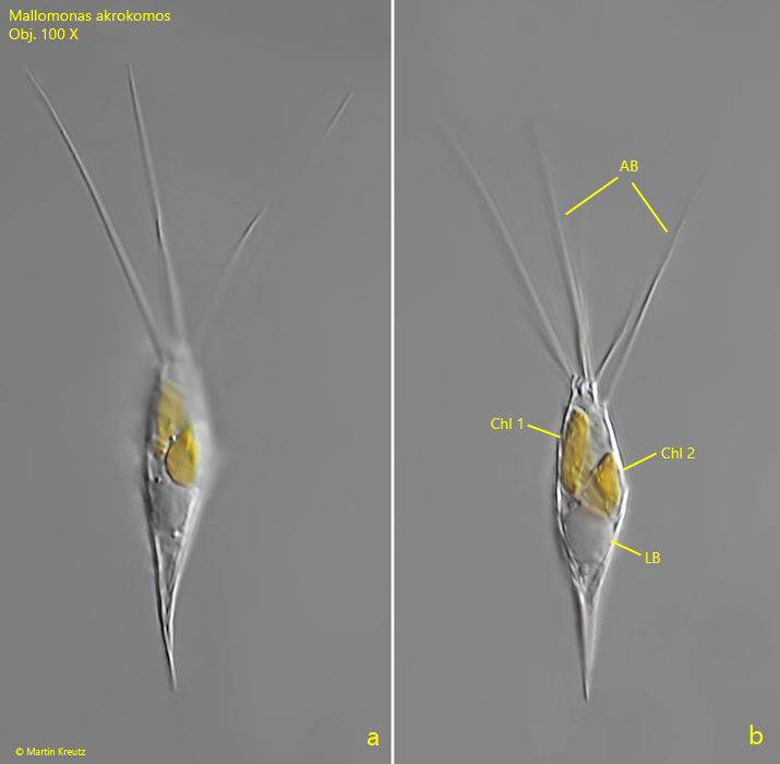
Fig. 1 a-b:Mallomonas akrokomos. L = 38 µm. Two focal planes of a freely swimming specimen. Note the tuft of long, apical brisles (AB) and the two chloroplasts (Chl 1, Chl 2). LB = leukosine body. Obj. 100 X.
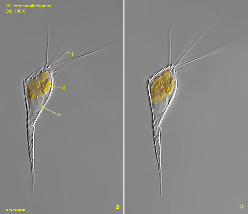
Fig. 2 a-b:Mallomonas akrokomos. L = 58 µm. A second freely swimming specimen. Chl = chloroplasts, F = flagellum, LB = leukosine body. Obj. 100 X.
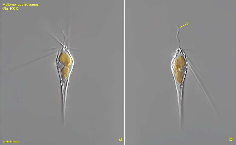
Fig. 3 a-b:Mallomonas akrokomos. L = 37 µm. A third freely swimming specimen. F = flagellum. Obj. 100 X.
Fig. 4:Mallomonas akrokomos. L = 41 µm. The delicate scales become visible in dead specimens. Obj. 100 X.
Fig. 5:Mallomonas akrokomos. A dead specimen that has previously formed a cyst (CY), which has also died. The cyst has a length of 14 µm. Obj. 100 X.