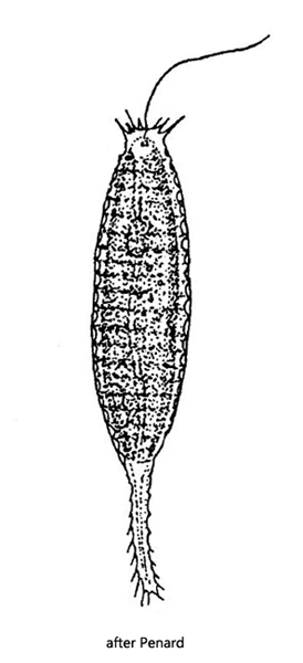
cells long elliptic, spindle shaped, posterior tail-like
length 70 – 100 µm
body without bristles, apical and caudal spines
scales concave and elliptical, with perforated rim, not covering each other
nucleus anterior
one apical flagellum
two elongated chromophores, golden-brown, yellowish or greenish
contractile vacuole consisting of 2-3 vesicles, anterior or basal
Mallomonas insignis
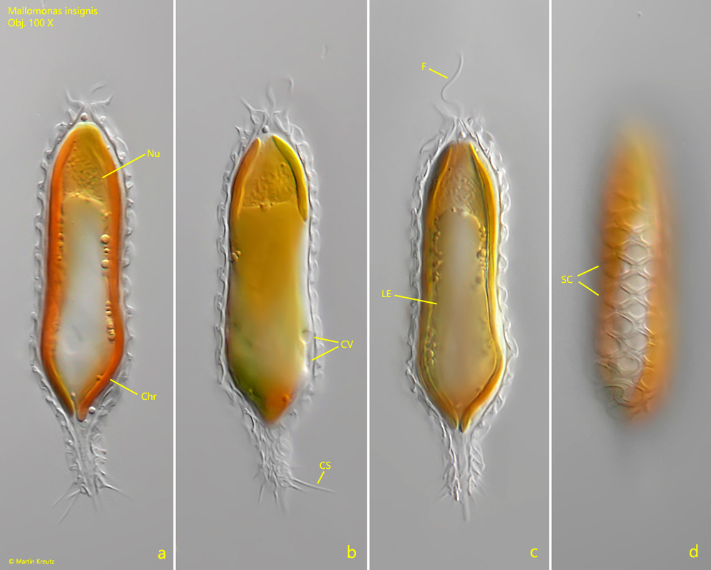
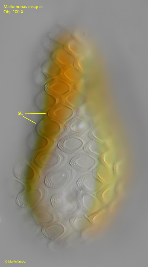
I regularly find Mallomonas insignis in plankton samples and in samples from the surface between floating plants or algae. At up to 100 µm, this chrysophyte is comparatively large and easily identified by the absence of spines. Instead, this alga has a tail-like appendage of silica scales with caudal spines (s. fig. 1b). The silica scales covering the body appear diamond-shaped at low magnifications and have a perforated rim, but this can only be seen at high magnifications (s. fig. 2).
Fig. 1 a-d:Mallomonas insignis. L = 75 µm. A freely swimming specimen. Chr = chromatophores, CS = caudal spines, CV = contractile vacuoles, F = flagellum, Nu = nucleus, LE = leucosin body, SC = scales. Obj. 100 X.
Fig. 2:Mallomonas insignis. The silica scales (SC) covering the cell in detail. Note the perforated rim of the scales. Obj. 100 X.