spherical nucleus (9–15 µm) with a central nucleolus
surface of nucleolus porous
numerous symbiotic algae scattered in cytoplasm
no crystals in cytoplasm
several contractile vacuoles
uroid rounded or bulbous
cell covered by inconspicuous coat
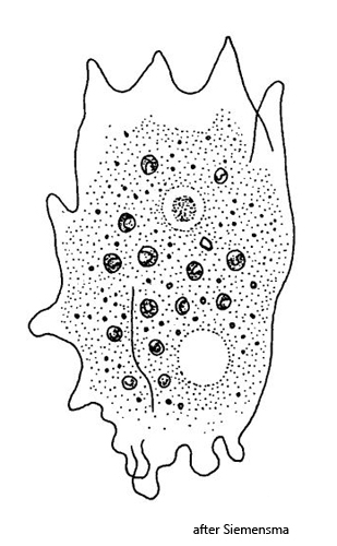
Mayorella viridis
Mayorella viridis is a very common naked amoeba in the Simmelried, which is easily recognized by its numerous symbiotic algae and the mammilliform pseudopodia. The nucleus is spherical with a central nucleolus. In my population the surface of the nucleolus was always porous (s. figs. 3 a and 4 b). The symbiotic algae are of the Chlorella type, have their own nucleus and a diameter of 4–6.5 µm (s. fig. 5). The cell is covered by a very thin cell coat, which can only be seen at high magnification (s. figs. 5 and 6). According to my measurements, it is 1.0-1.1 µm thick. The electron microscopical fine structure of the cell coat was investigated by Cann (1981). It is composed of fibrils that are arranged parallel and perpendicular to the cell surface.
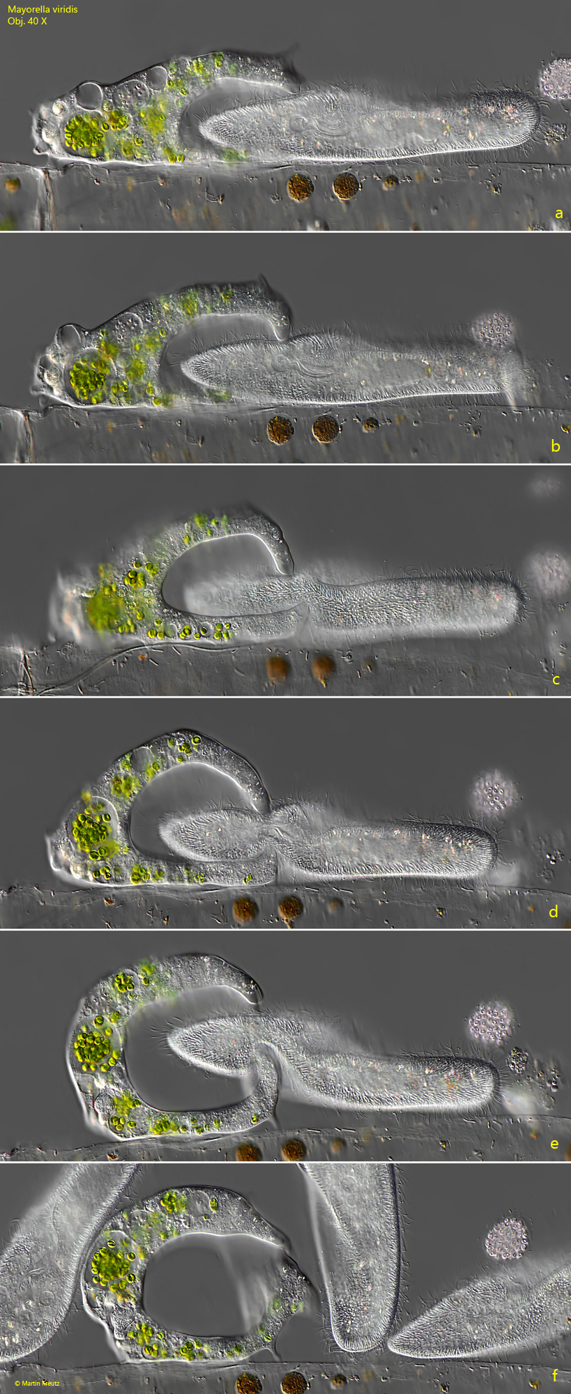
In the food vacuoles of Mayorella viridis I mostly found colony-forming bacteria, small algae, diatoms but also euglenids (e.g. Trachelomonas). In one case I was even able to observe an attack on Paramecium caudatum, but this was unsuccessful (s. fig. 7 a-f).
In February 2010, I found a specimen with an eneormously enlarged nucleus (s. fig. 8 a-b). The nucleus contained clumpy structures. This could be an enlarged nucleolus or a parasite has infested the nucleus.
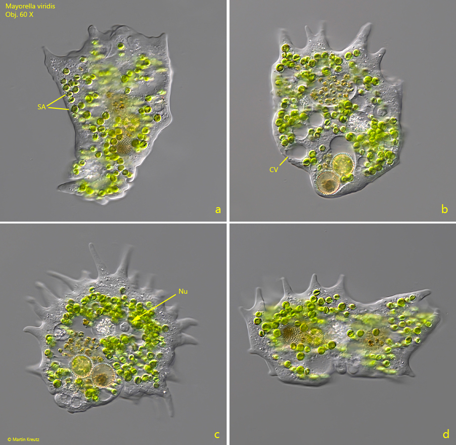
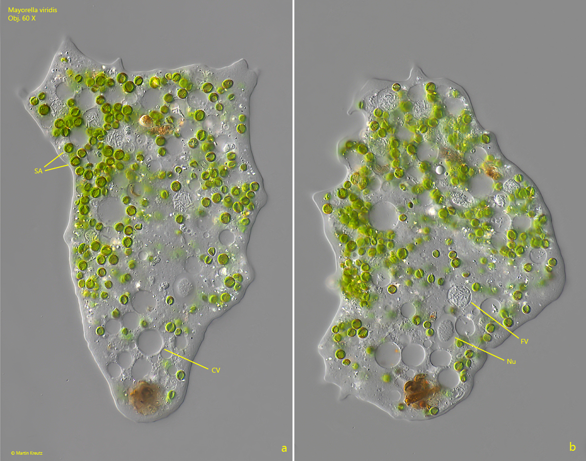
Fig. 1 a-d:Mayorella viridis. L = 164 µm. A free-flowing specimen. CV = contractile vacuole, Nu = nucleus, SA = symbiotic algae. Obj. 60 X.
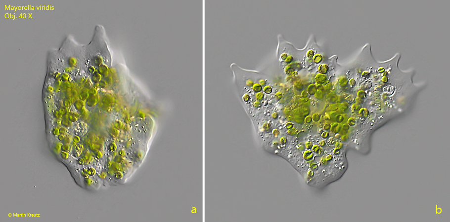
Fig. 2 a-b:Mayorella viridis. L = 135 µm. A second free-flowing specimen. Obj. 40 X.
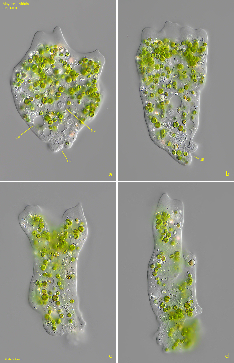
Fig. 3 a-d:Mayorella viridis. L = 135 µm. The same specimen as shown in fig. 2 a-b. CV = contractile vacuole, NU = nucleus, UR = uroid. Obj. 60 X.
Fig. 4 a-b:Mayorella viridis. L = 135 µm. The slightly squashed specimen as shown in fig. 2 a-b. CV = contractile vacuole, FV = food vacuole, NU = nucleus, SA = symbiotic algae. Obj. 60 X.
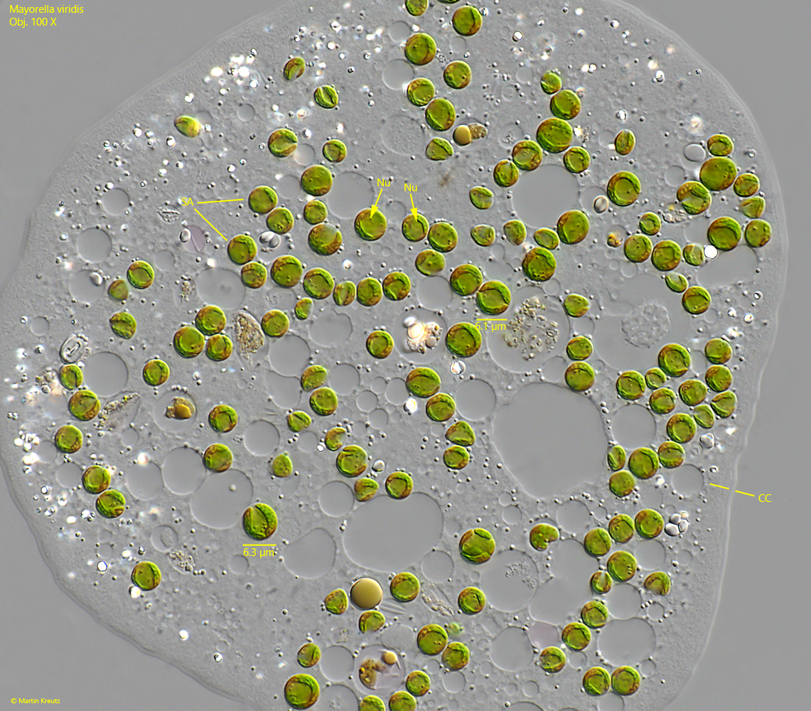
Fig. 5:Mayorella viridis. The symbiotic algae (SA) in a strongly squashed specimen. The algae are from the Chlorella type with a nucleus (Nu) and a diameter of 4–6.5 µm. Note the thin cell coat (CC). Obj. 100 X.
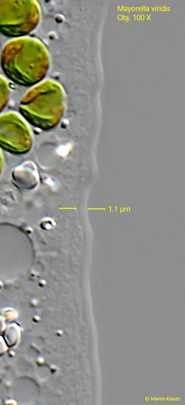
Fig. 6:Mayorella viridis. The inconspicuous cell coat with a thickness of 1.1 µm. Obj. 100 X.
Fig. 7 a-f:Mayorella viridis. A specimen tries to attack Paramecium caudatum. Obj. 40 X.
Fig. 8 a-b:Mayorella viridis. In this specimen the nucleus (Nu) seems to be enourmously enlarged and filled with a clumpy nucleolus (Nuc?) or the nucleus is infested by an unidentified parasite. FV = food vacuole. Obj. 100 X.