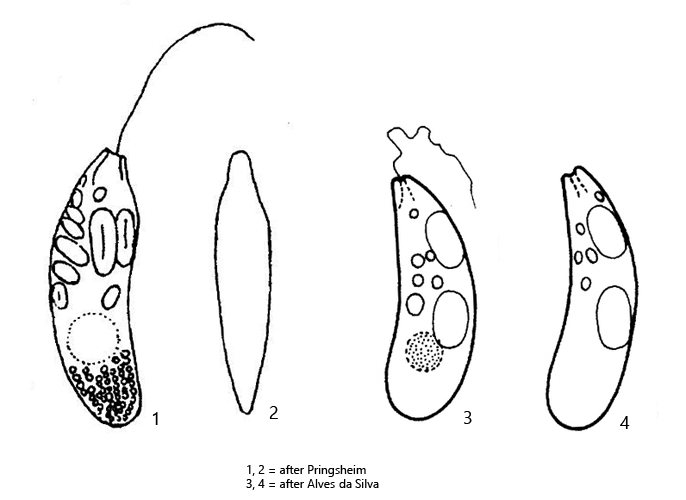
anterior end slightly tapered and transversely blunted
posterior end broadly rounded
pellicle with longitudinal striation
one flagellum, not reaching body length
nucleus globular below mid-body with central nucleolus
large paramylon grains in anterior half of cell
aggregation of small paramylon granules in posterior half
Menoidium obtusum
Menoidium obtusum is by far the most common species of the genus Menoidium in the Simmelried. There it sometimes emerges in enormous masses, with thousands of specimens per milliliter. However, until 2017, this species was completely absent from Simmelried. After that, the population has grew steadily and reached its current strength around 2019.
Menoidium obtusum can be easily identified by its somewhat clumsy shape in lateral view. The anterior end is not snout-like extended as in Menoidium pellucidum, but obliquely truncated. The distribution of the paramylon grains is typical of Menoidium. The anterior half contains the large paramylon grains, while the posterior half often contains a cluster of small paramylon grains. I also found transparent specimens that lacked large paramylon grains entirely (s. fig. 2 a-b). In these specimens, the nucleus was clearly visible, which is usually obscured by the paramylon grains (s. fig. 2 a). In addition, the delicate, longitudinal striation of the pellicle described by Pringsheim was visible in these transparent specimens (s. fig. 2 a). The specimens in my population were consistently about 50 µm long, with some specimens as long as 58 µm. This is longer than indicated by Pringsheim (30–37 µm). Menoidium obtusum was also described by Alves-da Silva and Friedrich (2009). These authors give a length of 41–42 µm. Due to the few descriptions of Menoidium obtusum, the variability of the body length is obviously not sufficiently evaluated.
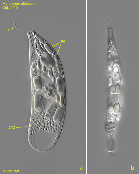
Fig. 1 a-b:Menoidium obtusum. L = 53 µm. A freely swimming specimen from left and from the narrow side. APG = aggregation of small paramylon grains, F = flagellum, PG = paramylon grains. Obj. 100 X.
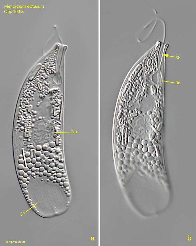
Fig. 2 a-b:Menoidium obtusum. L = 53 µm. A second specimen without large paramylon grains. Note the longitudinal striation of the pellicle (SP) and the short, secondary flagellum (SF) in the reservoir (Re). Nu = nucleus. Obj. 100 X.
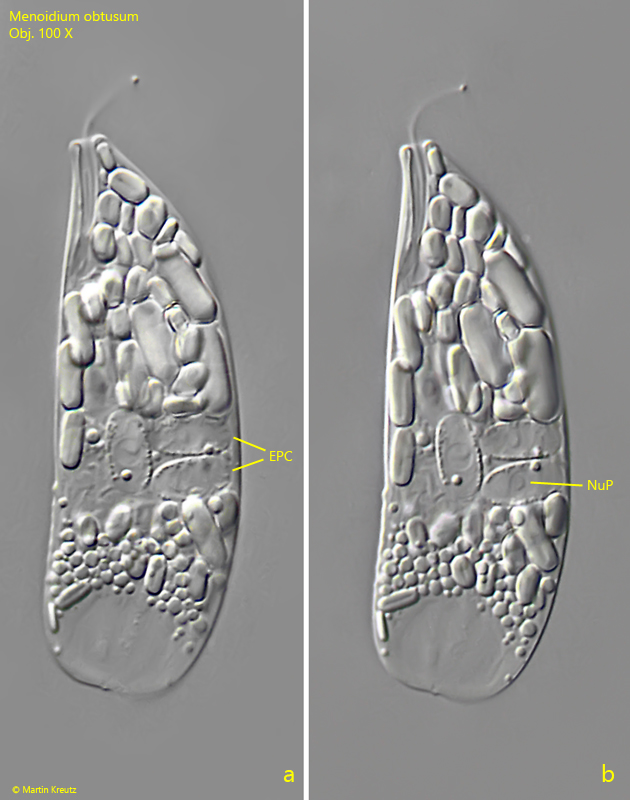
In my population I could find many specimens which were obviously infested by endoparasitic cells. Around the nucleus of these specimens, oval, transparent cells with a length of 7–8 µm were present (s. fig. 3 a-b). Each of these endoparasitic cells had its own nucleus. Possibly, this is an endoparasitic fungus. I could not observe developmental stages of this endoparasite.
Fig. 3 a-b:Menoidium obtusum. L = 49 µm. A specimen infested by endoparasitic cells (EPC). In each of these endoparasitic cells a separate nucleus is visible (NuP). The length of the endoparasitic cells is 7–8 µm. Obj. 100 X.
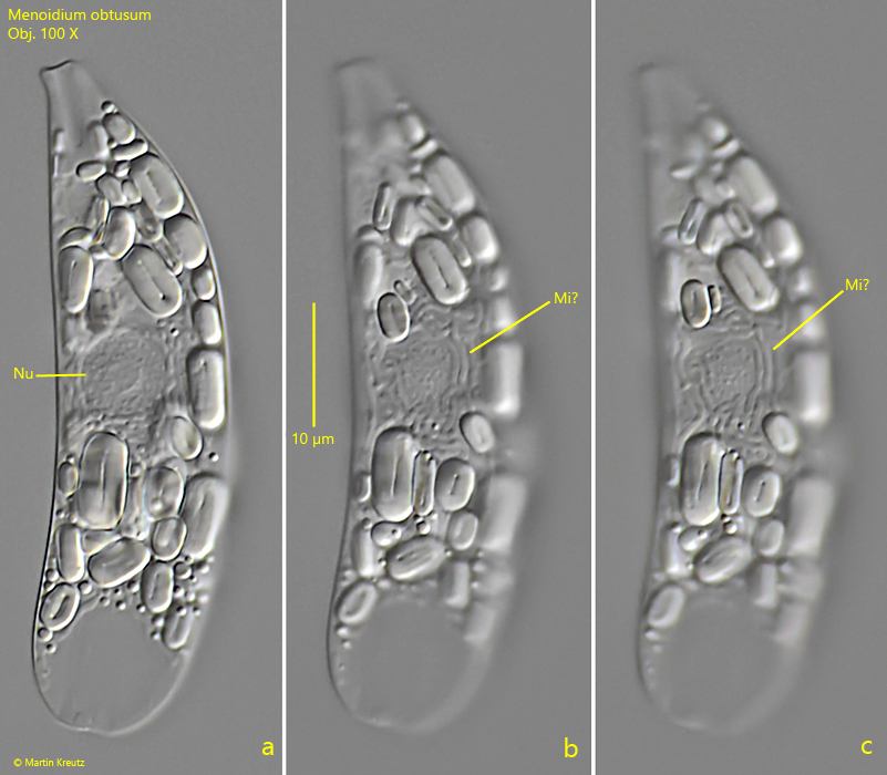
In other specimens of Menoidium obtusum I could see worm-like structures in the area around the nucleus (s. fig. 4 a-c). The nature of these structures is unknown to me. Possibly they are mitochondria, which sometimes can also have a worm-like appearance.
Fig. 4 a-c:Menoidium obtusum. L = 56 µm. A specimen with worm-like structures in the area around the nucleus. Possibly these are mitochondria (Mi?). Nu = nucleus. Obj. 100 X.