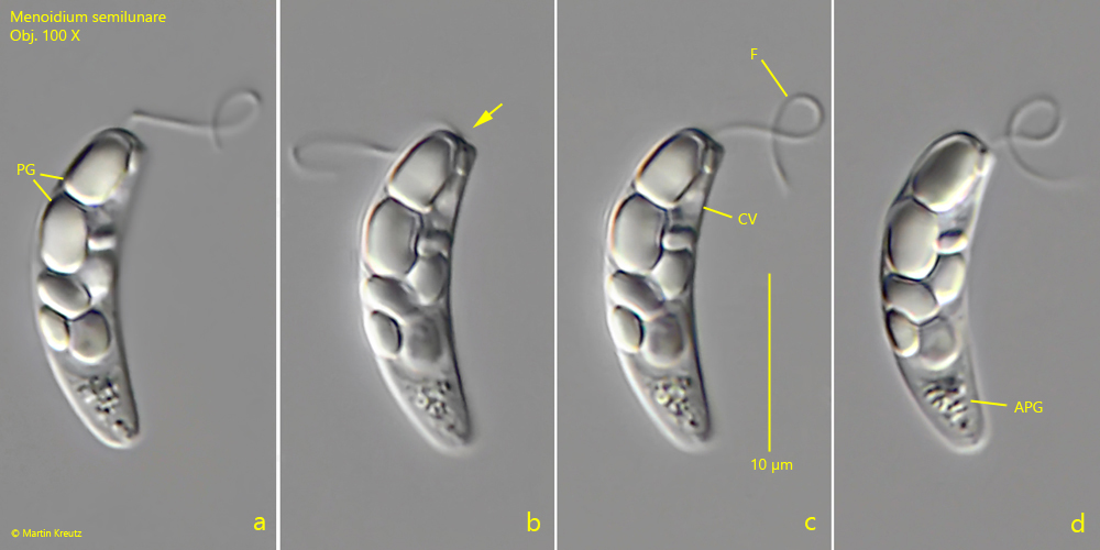
nucleus globular below mid-body with central nucleolus
large paramylon grains (up to 8 µm) in anterior half of cell
aggregation of small paramylon granules in posterior third
Menoidium semilunare
I found Menoidium semilunare in October 2021 in the Simmelried. Except for the two specimens shown below, I have no other records of this species so far.
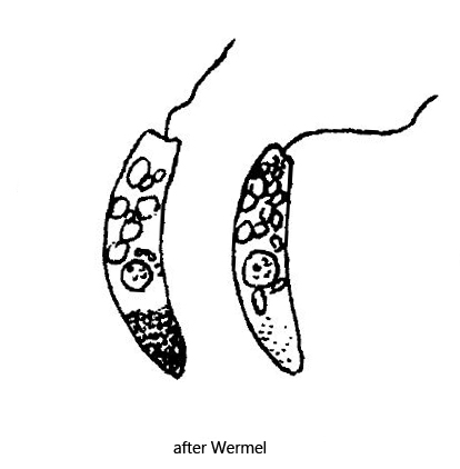
Of Menoidium semilunare apparently only the original description by Wermel is available, who found the species in 1924 in a bog near village Lutzino in Russia. Therefore the species is considered uncertain, since it is still insufficiently described. However, the specimens of my population agree very well with the description and drawings of Wermel, except for the length. Wermel gives 30 – 35 µm, while my two specimens were 19 µm long. However, since there are no other data about this species except the description of Wermel, the usual variability range for the length is still insufficiently known. Since all other characteristics of my specimens agree with the description of Wermel, I consider the identity to be proven.
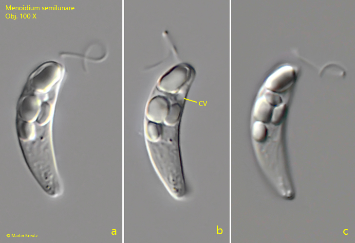
The body of Menoidium semilunare is curved and laterally flattened, like that of all representatives of the genus. The pellicle is smooth, as described by Wermel (s. fig. 2 c). In the anterior half of the body are large paramylon granules of broad oval shape, some even nearly rectangular. In my specimens they had a maximum length of 4.5 µm (Wermel gives max. 8 µm). In the posterior third of the body there is an accumulation of very small paramylon grains, the amount of which can obviously vary from specimen to specimen. The flagellum, according to my observations, is not quite as long as the body (s. fig. 1 c) and the contractile vacuole seems to be adjacent to the reservoir (s. figs. 1 c and 2 b). The anterior end is obliquely truncate with a slight indentation and the posterior end is slightly tapered and rounded as described by Wermel.
Fig. 1 a-d:Menoidium semilunare. L = 19 µm. A freely swimming specimen from right. Note the blunt anterior end with a slight dent (arrow) and the aggregation of small paramylon granules (APG) in the posterior third. CV = contractile vacuole, F = flagellum, PG = paramylon grains. Obj. 100 X.
Fig. 2 a-c:Menoidium semilunare. L = 19 µm. A second freely swimming specimen from right. CV = contractile vacuole. Obj. 100 X.