cells spherical, ellipsoidal or hemispherical (after cell division), 3–6 µm
mucilage sheath distinct, following conturs of cells
content of cells homogeneous
color blueish-green, often pale, sometimes pinkish
Merismopedia glauca
I found Merismopedia glauca in October 2003 in the Simmelried. After that I have not registered any more finds of this species.
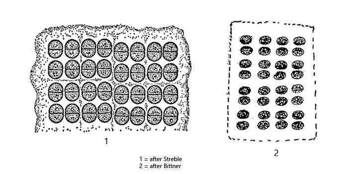
Merismopedia glauca is very similar to Merismopedia elegans. However, the colonies of Merismopedia glauca are much smaller and contain a maximum of 64 cells. In contrast, the tabular colonies of Merismopedia elegans can contain up to 4000 cells. In addition, the mucus envelope around the cells of Merismopedia glauca and surrounds the cells tightly, whereas the mucus envelope surrounding the colonies of Merismopedia elegans has a distance of about one cell diameter from the outer cells.
The color of Merismopedia glauca cells is apparently not uniform. It is said to be blueish-green and sometimes pinkish. The cells of my population were more olive green.
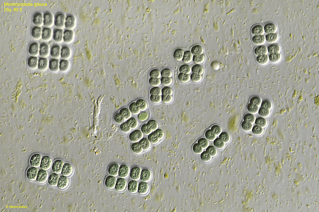
Fig. 1:Merismopediaglauca. Some colonies with 8–16 cells embedded in a layer of yellowish sulfur bacteria. Obj. 40 X.
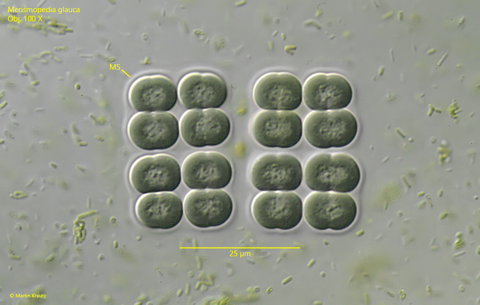
Fig. 2:Merismopediaglauca. A colony of 16 cells in detail. Each cell (in cell division) has a length of about 10 µm (= 5 µm per half-cell). Note the distinct mucilaginous sheath (MS) following the conturs of the cells. Obj. 100 X.