adoral zone running obliquely over ventral side before it bent to posterior end
mouth opening in mid-body
perizonal cilia long
macronucleus oval or ellipsoidal
contractile vacuole terminal
no elongated caudal cilia
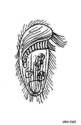
Metopus barbatus
So far I could find only some specimens of Metopus barbatus in February 2008. Also Kahl, who provides only a very short description, has found only a few specimens and describes this species as rare.
The body of Metopus barbatus is uniformly oval and not narrowed posteriorly as in Metopus setosus. In addition, this species lacks the long caudalcilia of Metopus setosus. However, I could observe slightly elongated caudalcilia in my specimen (s. fig. 1 d), but they by no means reach the length of the similar species Metopus setosus. The body of Metopus barbatus is also dorsal-ventrally flattened as described by Kahl (s. fig. 1 d). However, I could detect a slight concave depression of the ventral side (s. fig. 1 d), which Kahl does not mention. Otherwise, all visible features agree with the description by Kahl.
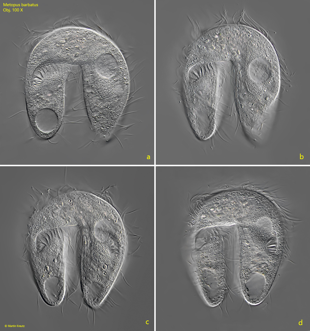
In the population of February 2008 I also found some specimens which were just in conjugation. In this case the specimens are connected via their apical domes to form a bridge of cytoplasm and not by the mouth openings as in most other ciliates. The mouth openings of the joined specimens point in opposite directions. This can be seen in the course of the adoral zones in fig. 2 c, which are mirror symmetrical. If the mouth openings would point in the same direction, the course of the two adoral zones would be congruent.
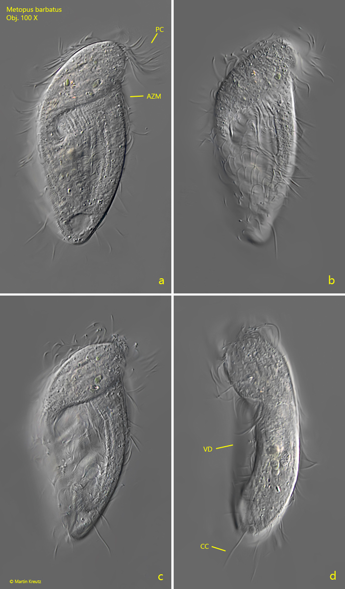
Fig. 1 a-d:Metopus barbatus. L = 73 µm. A freely swimming specimen from ventral (a-c) and from left (d). Note the concave depression of the ventral side (VD). At the posterior end slightly elongated caudal cilia (CC) are visible with a length of about 10 µm. AZM = adoral zone of membranelles, PC = perizonal cilia. Obj. 100 X.
Fig. 2 a-d:Metopus barbatus. L = 62 µm. Two freely swimming specimen in conjugation. Obj. 100 X.