body twisted, apical dome to the left, anterior end to the right
curved or almost U-shaped in ventral or dorsal view
length about 45 µm
adoral zone short, mouth opening in mid-body
perizonal ciliary row running in parallel to adoral zone
macronucleus oval, located in apical dome
contractile vacuole terminal
tuft of some elongated caudal cilia
Metopus convexus
The only description and drawing of Metopus convexus known to me is by Kahl and is very short. In my sampling sites Simmelried and Purren pond I find Metopus convexus rarely, but regularly. Although the species is small, it can be recognized by the typically curved body shape even at low and medium magnifications. Some specimens are almost U-shaped. Additionally, the two legs of the U (i.e. anterior and posterior end) are also twisted against each other. That means the body is curved and twisted. Examination of Metopus convexus is not easy, because in my experience the species is quite coverslip sensitive and denatures quickly when the layer thickness is reduced.
The apical dome of Metopus convexus is prominent and protrudes strongly. In my population the macronucleus was localized in the anterior half near the apical dome (s. figs 1e) or in mid-body (s. fig. 2 d-f). The spherical micronucleus (not mentioned by Kahl) is separated from the macroneucleus with a diameter of 4.5 µm (s. fig. 2 e). According to my observation, the adoral zone begins at the right side of the dome and extends to the ventral side above the cell equator, where the mouth opening is localized (s. fig. 1 a and 2 a). Above the adoral zone the perizonal ciliary row runs in parallel also to the mouth opening (s. fig. 3 a). At the posterior end, some elongated caudal cilia protrude in a fan shape (s. figs. 2 b, 2 d and 2 f). The contractile vacuole lies terminally. In the cytoplasm I could clearly recognize symbiotic bacteria (s. fig. 3 c). Kahl does not mention them, but many other metopid ciliates also have symbiotic bacteria.
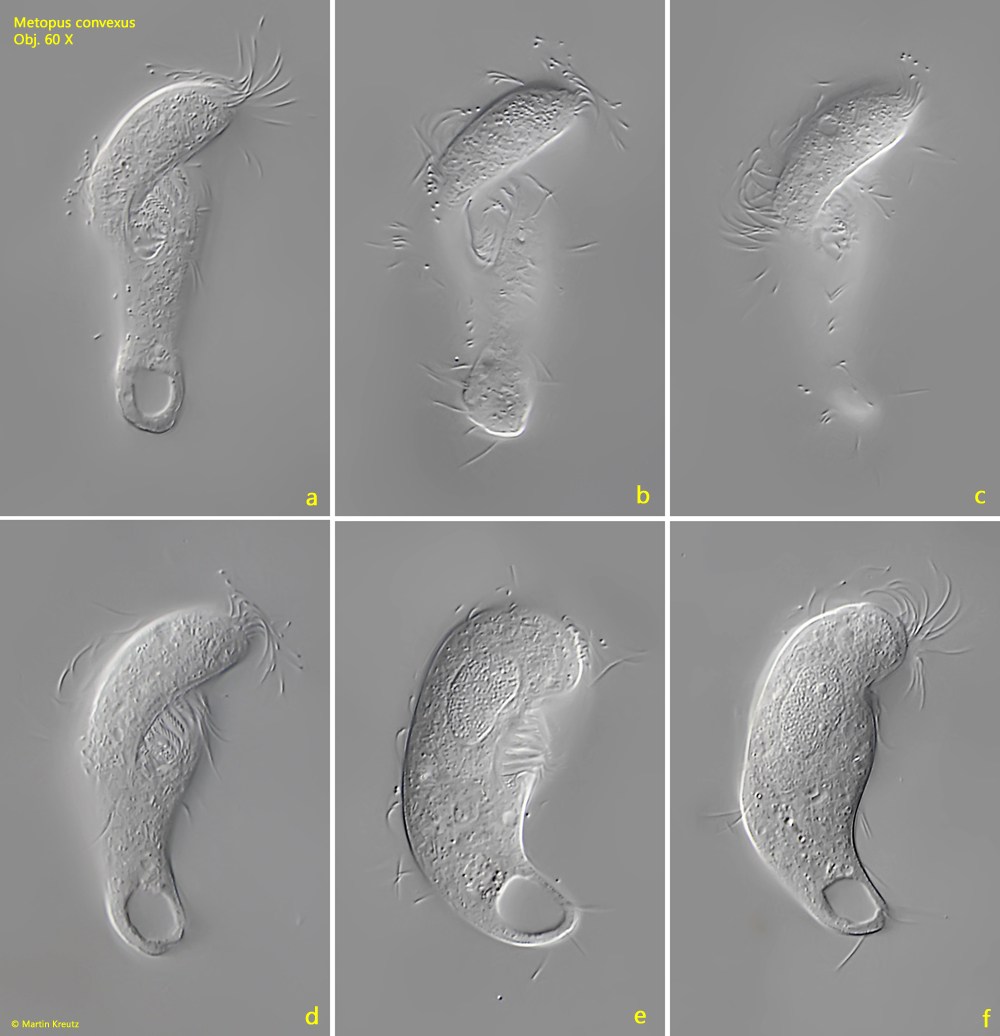
Fig. 1 a-f:Metopus convexus. L = 54 µm. A freely swimming specimen from ventral (a-d) and from right (e, f). Obj. 60 X.
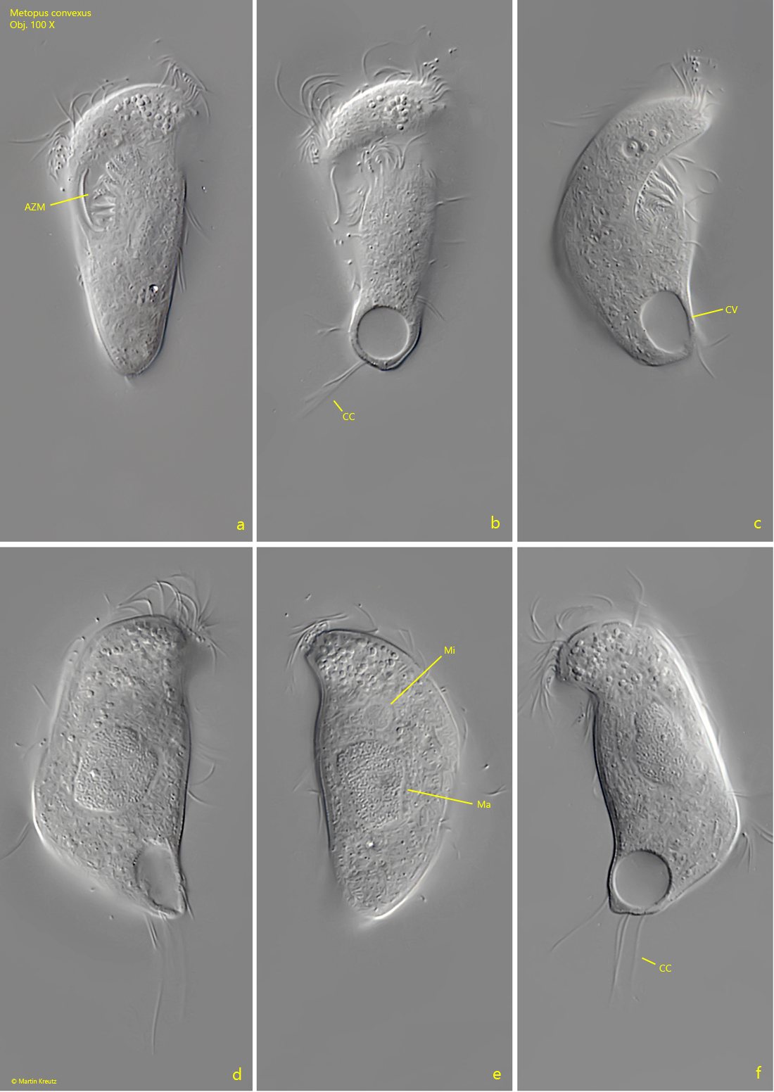
Fig. 2 a-f:Metopus convexus. L = 49 µm. A second freely swimming specimen from ventral (a, b), right (c, d) and left (e, f). Note the caudal cilia (CC) of the specimen. The spherical micronucleus (Mi) is separated from the macronucleus (Ma). AZM = adoral zone of membranelles. Obj. 100 X
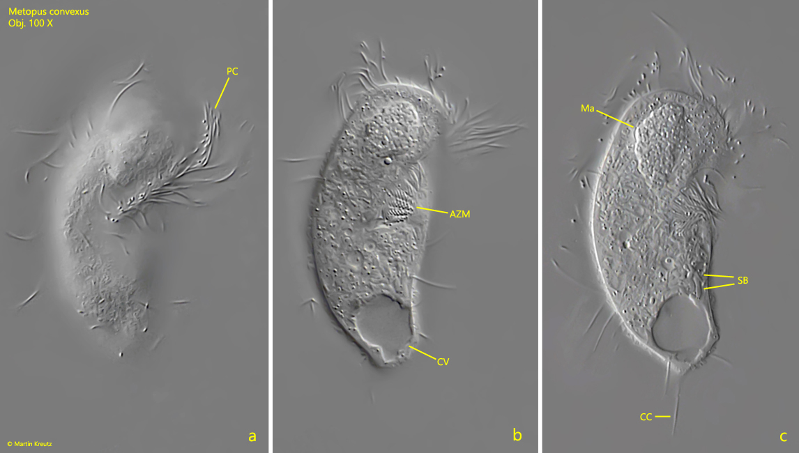
Fig. 3 a-c:Metopus convexus. L = 46 µm. Ventral view of slightly squashed specimen. Note the row of perizonal cilia (PC) running in parallel to the adoral zone of membranelles (AZM) to the mouth opening in mid-body. CC = caudal cilia, CV = contractile vacuole, Ma = macronucleus, SB = symbiotic bacteria in the cytoplasm. Obj. 100 X