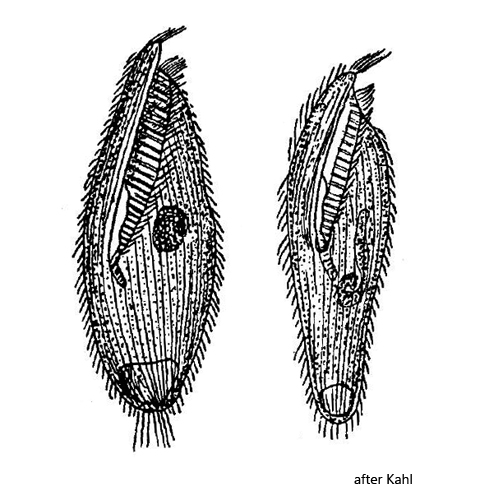
body slender oval, dorso-ventrally little flattened
length about 160 µm
ectoplasm brownish granular
perizonal cilia long
adoral zone runs obliquely over ventral side
membranelles of adoral zone high and densely arranged
mouth opening in posterior third
macronucleus rod-shaped, either entire or curled at end
contractile vacuole terminal
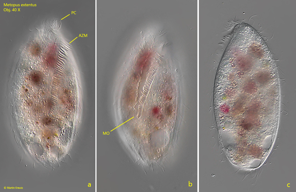
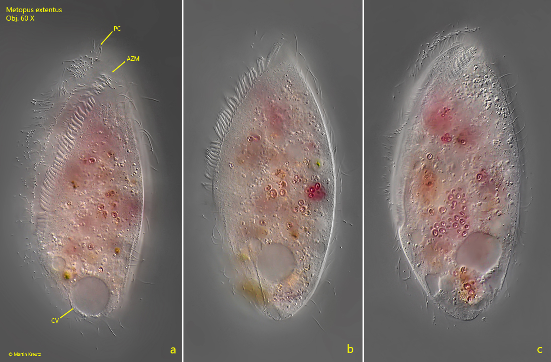
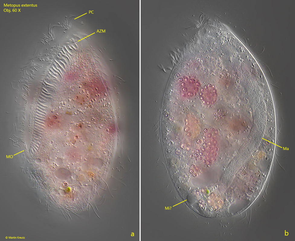
Metopus extentus
So far I could find only one specimen of Metopus extentus in February 2022 in the Purren pond. This fits the description of Kahl, who describes the species as “rare” which can be found in winter and spring.
Metopus extentus is easily recognized by its stately size of about 160 µm (my specimen was 143 µm long) and the adoral zone extending obliquely across the ventral side (s. figs. 1 a and 1b). The mouth opening is below the middle of the cell, at the border of the posterior third. I could not observe the brown granules in the ectoplasm described by Kahl, although the specimen appeared brownish at lower magnification (s. figs. 1 a-c). However, this was due to the contents of the food vacuoles. Unfortunately, my specimen was completely filled with food vacuoles containing mainly rhodobacteria in various stages of digestion. Thus, I could only clearly recognize the rod-shaped macronucleus (s. fig. 3b), which, according to Kahl, should sometimes be completely curled up or just one end. I could not clearly identify the micronucleus, since it could also be the optical section through a rolled-up end of the macronucleus (s. fig. 3 b).
In the literature and on the internet I often found the name “Metopus extensus”. However, Kahl has called this species Metopus extentus. I suspect a transcription error behind it.
Fig. 1 a-c:Metopus extentus. L = 143 µm. A freely swimming specimen from ventral (a, b) and from left (c). AZM = adoral zone of membranelles, MO = mouth opening, PC = perizonal cilia. Obj. 40 X.
Fig. 2 a-c:Metopus extentus. L = 143 µm. The same specimen shown in fig. 1 a-c from ventral (a) and from left (b, c). AZM = adoral zone of membranelles, CV = contractile vacuole, PC = perizonal cilia. Obj. 60 X.
Fig. 3 a-b:Metopus extentus. L = 143 µm. Focal plane on the adoral zone (a) and on the rod-shaped macronucleus (b) of the slightly squashed specimen shown in fig. 1 a-c. Near the posterior end likely the micronucleus (Mi?) is visible or the curled end of the macronucleus. AZM = adoral zone of membranelles, Ma = macronucleus, MO = mouth opening, PC = perizonal cilia. Obj. 60 X.