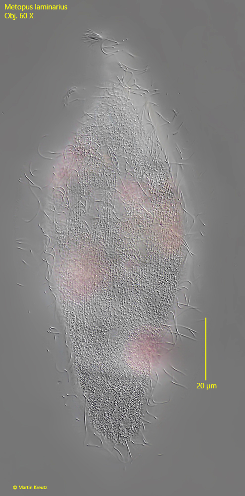
body cylindrical, sometimes irregular due to food content
posterior end transversely truncated
length 200–260 µm
always with pink rhodobacteria in food vacuoles
cytoplasm sometimes yellowish
anterior end flattened and spirally elongated
adoral zone runs along the spiral extension to mouth opening
mouth opening in anterior third
contractile vacuole terminal, often dilated towards anterior end
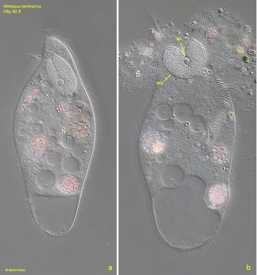
macronucleus sausage-shaped in anterior end, often curved into a horseshoe shape
micronucleus spherical, adjacent to the macronucleus or enclosed by it in a U-shape
very fine, long caudal cilia (hard to see)
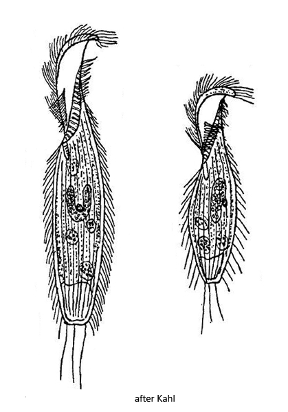
Metopus laminarius
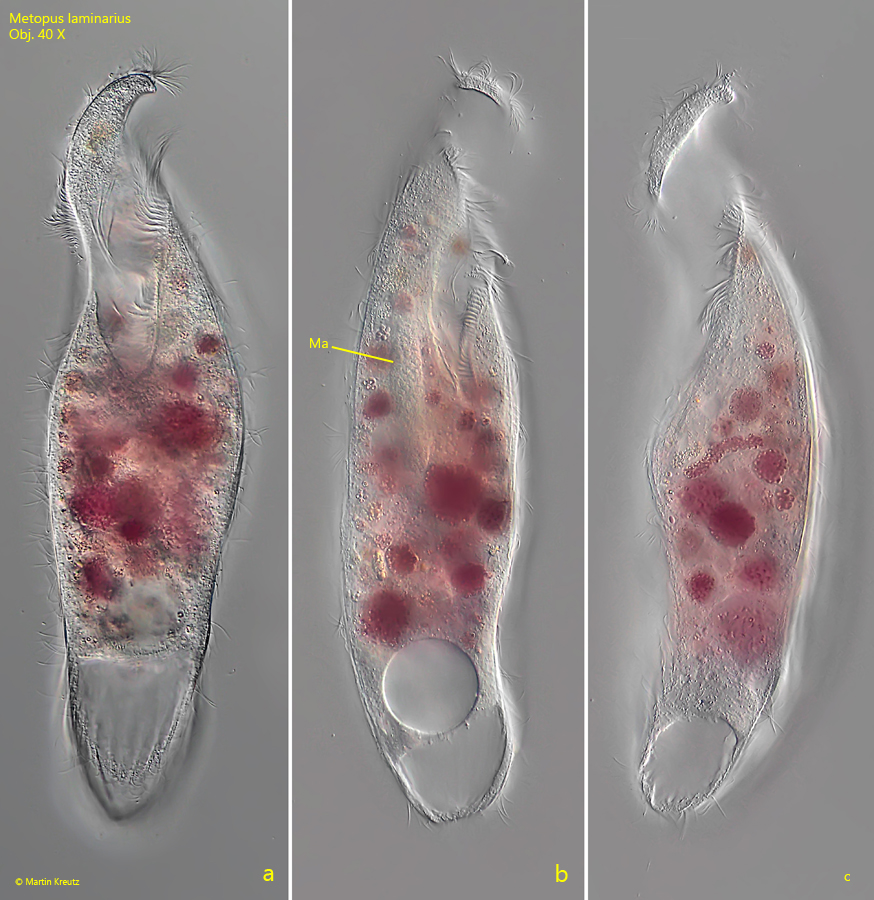
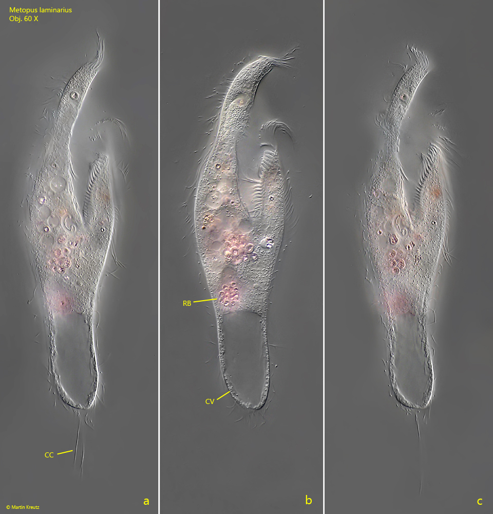
In my sampling sites Purren pond and Simmelried I find Metopus laminarius frequently and regularly. It is one of the largest species of the genus Metopus. Kahl gives a range of 200–260 µm for the body length and distinguishes a variety “minor” (s. right drawing above) with a length of about 150 µm. In my population I could find specimens with lengths between 170 – 320 µm. The body size seems to depend strongly on the number of food vacuoles in the body. Well fed specimens were larger. Therefore, in my opinion, the variety “minor” is not justified, since it is only a sign of nutritional status. As described by Kahl, the food vacuoles of Metopus laminarius are mainly filled with pink rhodobacteria (s. fig. 2b). The cytoplasm often appeared faintly yellowish. The anterior end is flattened and twisted in a spiral running in clockwise direction. The adoral zone follows this spiral and lowers ventrally into the mouth opening (s. fig. 3a). The long caudal cilia, described and drawn by Kahl, are hard to see. Only in one specimen I could document them (s. figs. 2 a and 2 c). In other specimens I sometimes could not see them at all. Possibly they are shed quickly. Metopus laminarius lacks a fringe of extrusomes under the pellicle, but it appears finely granulated (s. fig. 5).
Fig. 1 a-c:Metopus laminarius. L = 312 µm. A freely swimming specimen from ventral (a, b) and from left (c). Note the spirally twisted anterior end. Ma = macronucleus. Obj. 40 X.
Fig. 2 a-c:Metopus laminarius. L = 180 µm. A freely swimming specimen from ventral. Note the long caudal cilia (CC). RB = food vacuoles filled with ingested rhodobacteria. Obj. 60 X.
Fig. 3 a-b:Metopus laminarius. L = 280 µm. The slightly squashed specimen from ventral (a) and from right (b). AZM = adoral zone of membranelles, Ma = macronucleus, Mi = micronucleus. Obj. 60 X.
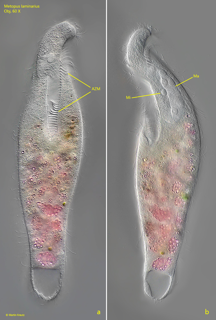
Fig. 4 a-b:Metopus laminarius. A specimen with a horseshoe shaped macronucleus (Ma) during continuous reduction of the layer thickness. The macronucleus encloses the spherical micronucleus (Mi). Obj. 60 X.
Fig. 5:Metopus laminarius. Focal plane on the granulated pellicle in a squashed specimen. Obj. 60 X.