body elongated oval, posterior half slightly tapered, dorso-ventrally slightly flattened
apical end elongated to a trunk-shaped organelle with pointed distal end, 35–50 µm long
trunk-shaped organelle towards the left side of the body
length 100–130 µm
adoral zone reaches the cell equator
pharynx slightly below cell equator, bent left
macronucleus spherical or ellipsoidal with one adjacent, spherical micronucleus
fringe of about 1 µm long extrusomes beneath the pellicle
contractile vacuole terminal
caudal cilia 10–30 µm long
Metopus nasutus
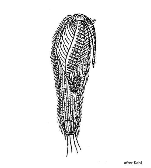
Metopus nasutus is one of the most common metopid ciliates in the Simmelried and the Purren pond. Especially in the Purren pond this remarkably shaped Metopus species can be found reliably. Metopus nasutus is easily recognized by a trunk-shaped organelle located at the anterior end. This organelle of unknown function makes the species unmistakable.
In my population I could find specimens with body lengths between 90–170 µm, which extends the range given by Kahl (100–150 µm). The trunk-shaped organelle is passively dragged along when swimming and lies against the left side of the body, as Kahl describes it (s. fig. 7 a-b). Only once could I observe the organelle to be stretched forward (s. fig. 3 a-d). In this specimen the trunk-shaped organelle was also unusually long with 74 µm. Kahl gives a length of 10–30 µm. On which factors the length of the organelle depends is unknown. In any case, it can melt under mechanical stress (coverslip pressure).
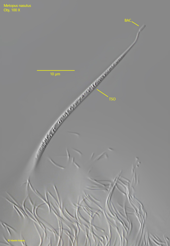
At high magnification, many vesicles and granules can be seen in the trunk-shaped organelle (s. fig. 6). In one case, I was able to find an attached bacterium at the tip of the organelle (s. fig. 6). Possibly this is a coincidental constellation, however it made me think of the possibility that the trunk-shaped organelle may be used like a glue rod to collect bacteria and other food particles. This theory is supported by the observation that in some free-swimming specimens I had the impression that the trunk was sometimes located between the perizonal cilia and the adoral membranelles in the direction of the mouth opening (s. figs. 8 a-b and fig. 9). Possibly bacteria or other food particles are “brushed off” between the perizonal cilia and the adoral membranelles during this behavior.
Beneath the pellicle of Metopus nasutus a fringe of extrusomes about 1 µm long is located (s. fig. 5a), not mentioned by Kahl. Many symbiotic bacteria are visible in the cytoplasm (s. fig. 5 a), especially in the area around the macronucleus. In the food vacuoles I found colorless bacteria, green-yellow sulfur bacteria, pink rhodobacteria and small green algae.
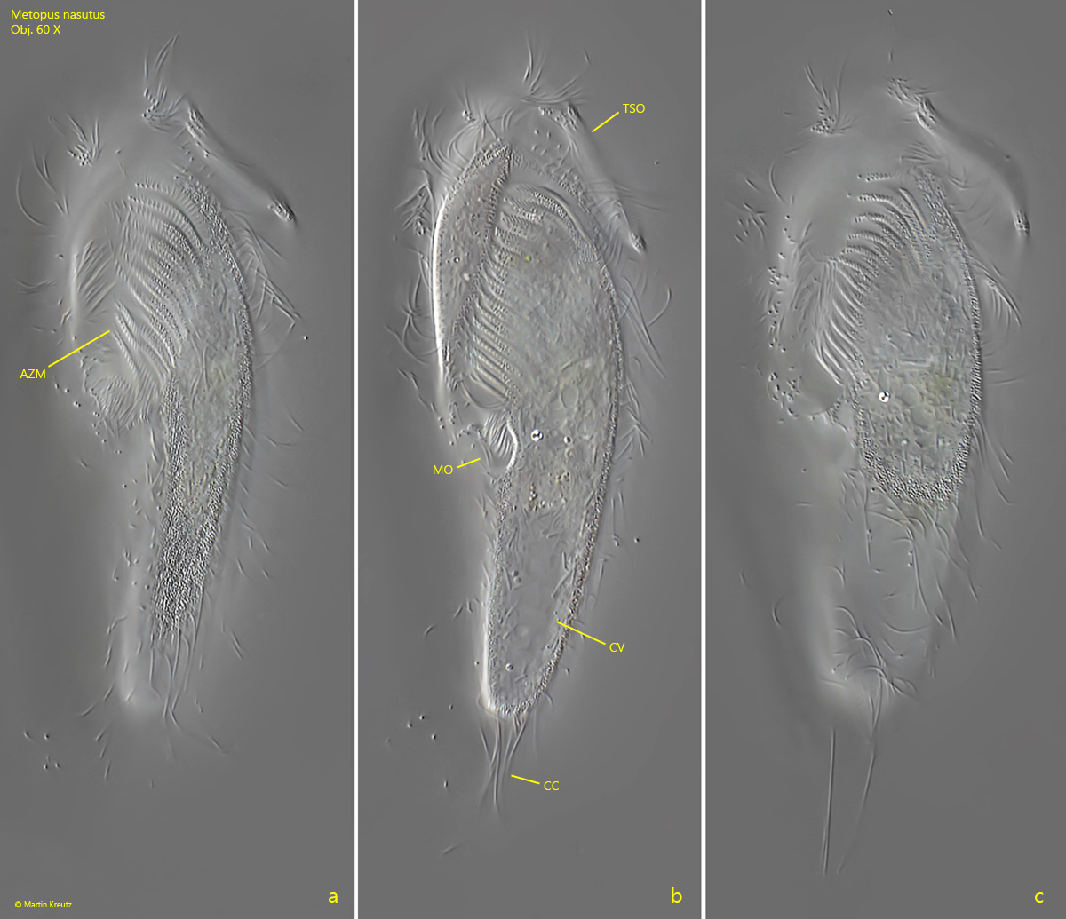
Fig. 1 a-c:Metopus nasutus. L = 165 µm. Ventral view of a freely swimming specimen. Note the trunk-shaped organelle (TSO) at apical end. AZM = adoral zone of membranelles, CC = caudal cilia, CV = contractile vacuole, MO = mouth opening. Obj. 60 X.
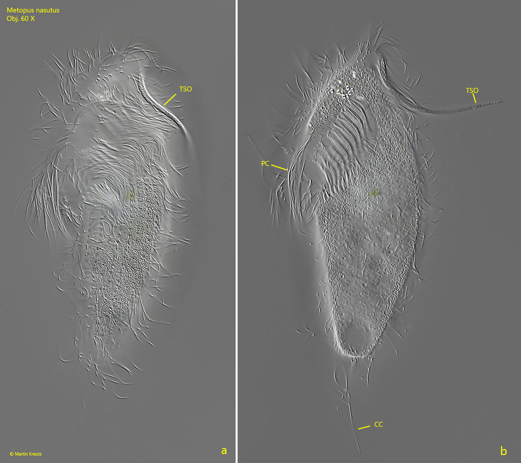
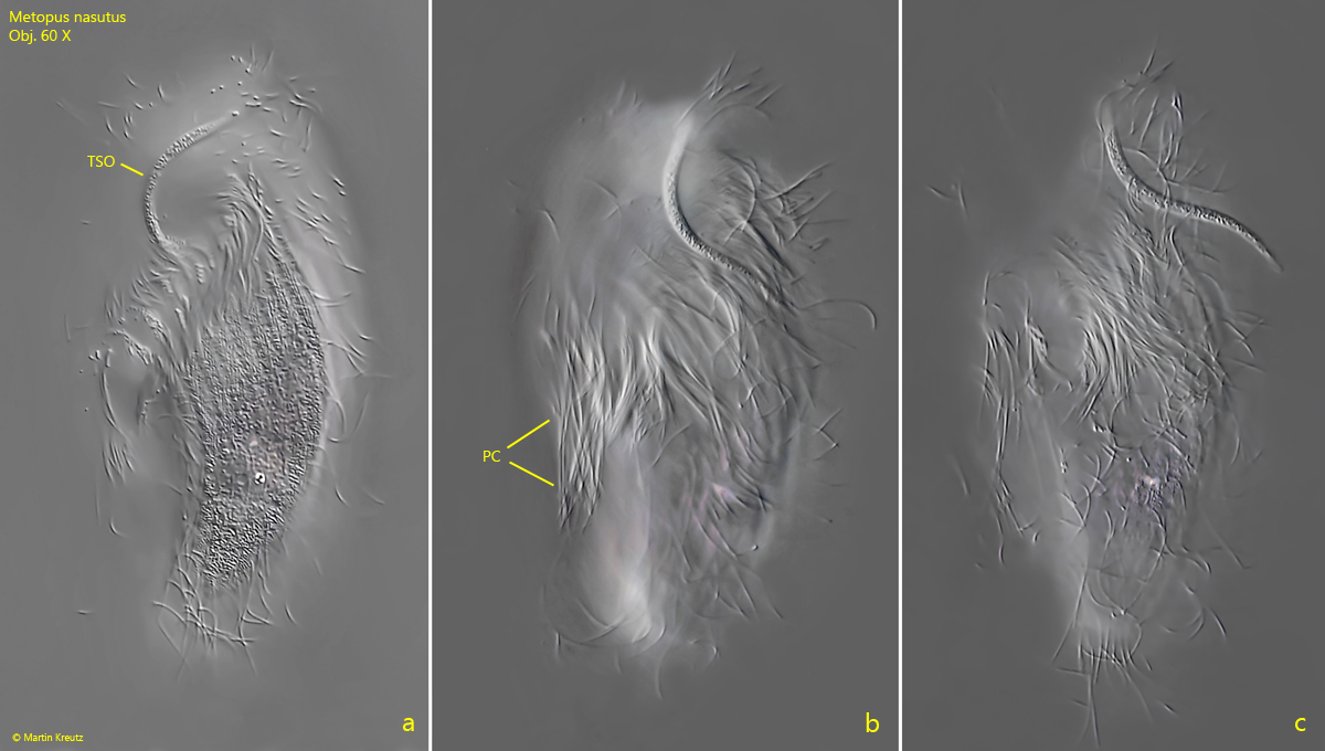
Fig. 2 a-b:Metopus nasutus. L = 115 µm. Ventral view of a seond, freely swimming specimen. CC = caudal cilia, PC = perizonal cilia, TSO = trunk-shaped organelle. Obj. 60 X.
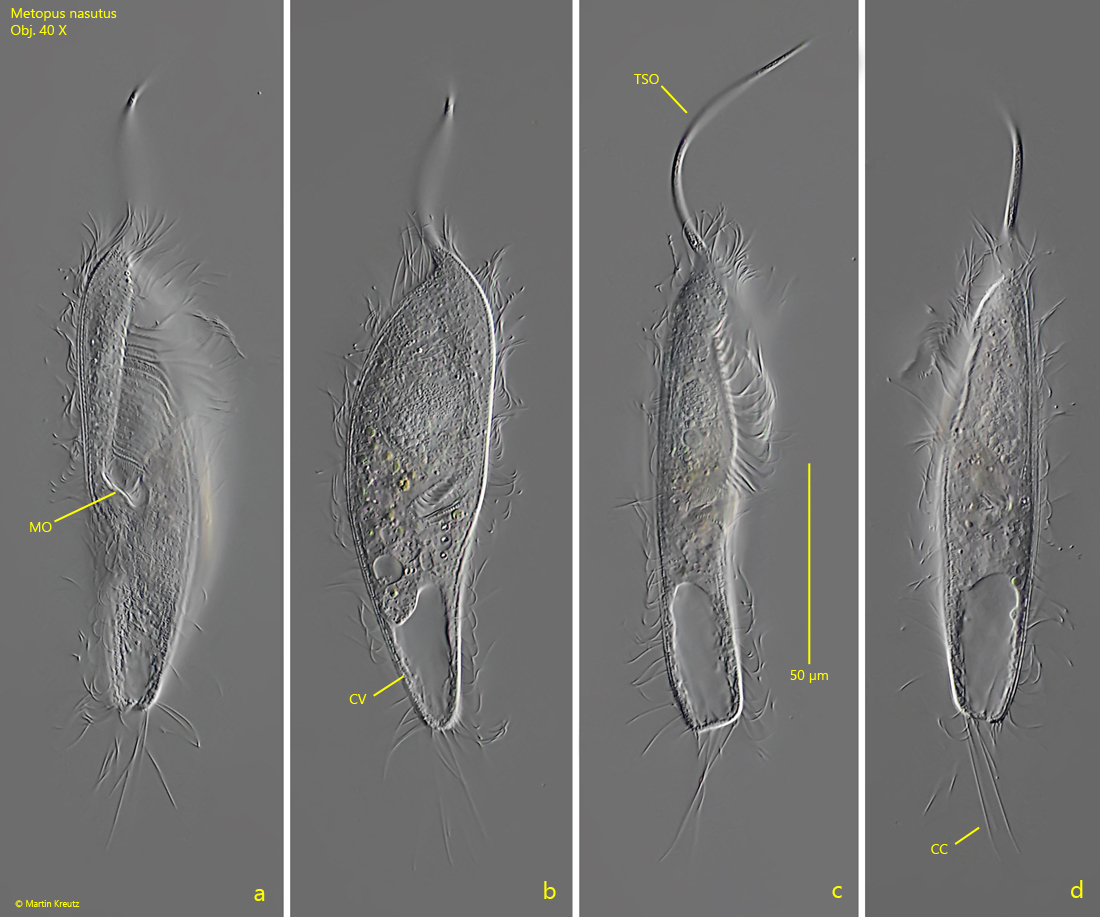
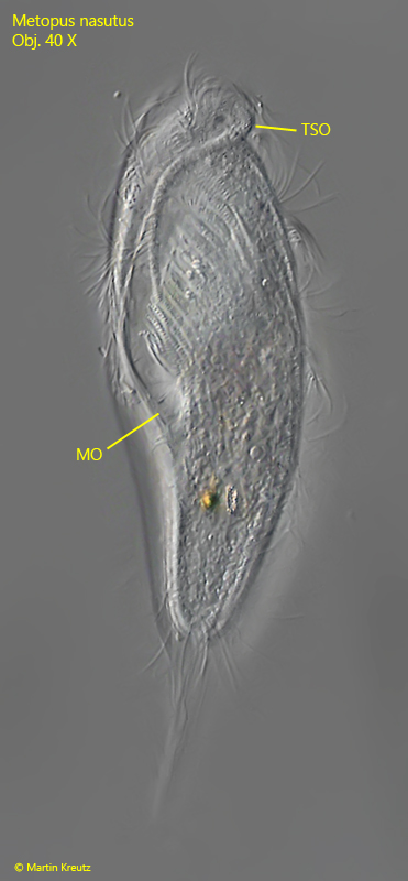
Fig. 3 a-d:Metopus nasutus. L = 122 µm (without trunk shaped organelle). A freely swimming specimen with an exceptionally long trunk-shaped organelle (L = 74 µm) that is extended forward in the direction of swimming. CC = caudal cilia, CV = contractile vacuole, MO = mouth opening. Obj. 40 X.
Fig. 4 a-c:Metopus nasutus. L = 92 µm. Focal plane on the trunk-shaped organelle (TSO) in an freely swimming specimen. Note the long perizonal cilia (PC). Obj. 60 X.
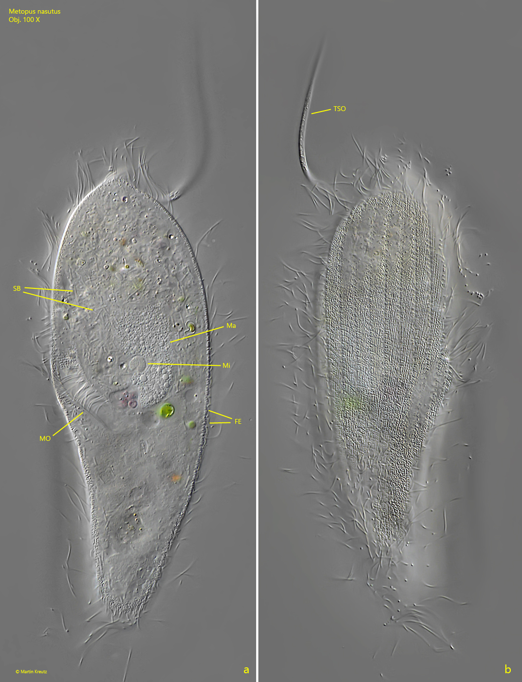
Fig. 5 a-b:Metopus nasutus. A sligthly squashed specimen from ventral (a) and dorsal (b). Note the symbiotic bacteria in the cytoplasm (SB) and the distinct striation of the granulated pellicle (b). Beneath the pellicle a fringe of about 1 µm long extrusomes is visible (FE): Ma = macronucleus, Mi = micronucleus, MO = mouth opening, TSO = trunk-shaped organelle. Obj. 100 X.
Fig. 6:Metopus nasutus. The trunk-shaped organelle (TSO) in detail. In the trunk many vesicles and granules are located. At the tip of the trunk an adherent bacterium is visible (BAC). This may be there by chance or it may be an indication that the trunk has a function similar to a glue rod to collect bacteria. Obj. 100 X.
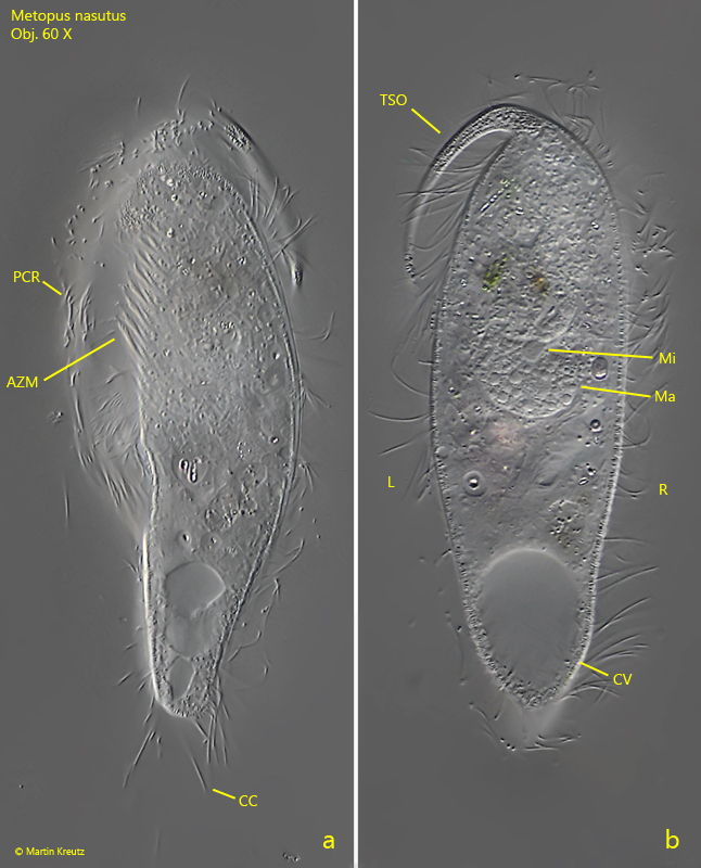
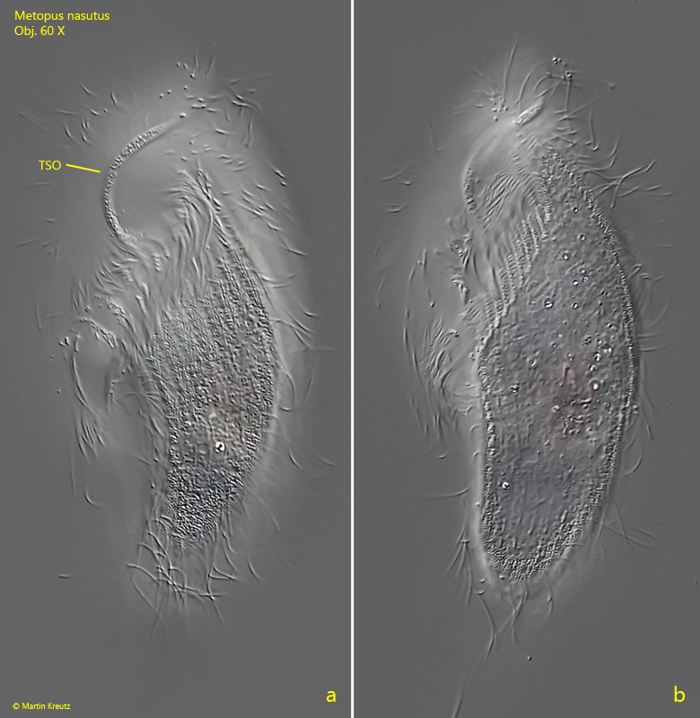
Fig. 7 a-b:Metopus nasutus. L = 115 µm. A freely swimming specimen from left (a) and from dorsal (b). The trunk-shaped organelle (TSO) lies passively on the left side of the body when swimming. AZM = adoral zone of membranelles, CC = caudal cilia, CV = contractile vacuole, L/R = left/right, Ma = macronucleus, Mi = micronucleus, PCR = perizonal cilia row. Obj. 60 X.
Fig. 8 a-b:Metopus nasutus. L = 96 µm. A freely swimming specimen from left with the trunk-shaped organelle (TSO) bent in the direction of the mouth opening. Obj. 60 X.
Fig. 9:Metopus nasutus. L = 148 µm. In this freely swimming specimen the trunk-shaped organelle (TSO) seems to located between the perizonal cilia row and the adoral zone, bent in direction of the mouth opening (MO). Obj. 40 X.