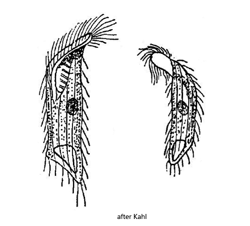
body cylindrical, parallel-sided, ventrally curved
posterior end obliquely truncated
ventral side keeled
length 50–60 µm
adoral zone runs obliquely to the mid-body
cilia loosely arranged, not elongated
macronucleus spherical
contractile vacuole terminal in ventral keel
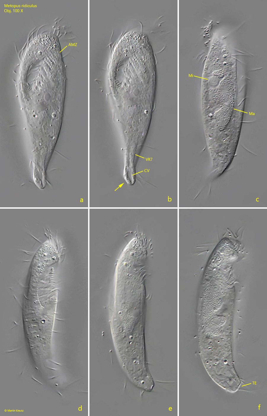
Metopus ridiculus
Apart from the very short description by Kahl I have not found any further information about Metopus ridiculus. He describes the species as “not common” and from his description it seems that he could not examine Metopus ridiculus thorougly (likely due to lack of specimens), as he writes for example “(adoral) zone probably up to half”. I have found only two specimens Metopus ridiculus so far. The first in the Mainau pond in October 2019 (s. fig. 1 a-f) and the second in the Simmelried in December 2022 (s. fig. 2 a-d). Kahl highlights the truncated posterior end, which he drew in ventral view (s. drawings above). In my first found specimen (s. fig. 1 a-f) such a truncated end is only slightly pronounced in ventral view (s. fig. 1b) but clearly visible in lateral view (s. fig. 1 f). The body of this specimen is not ventrally curved throughout, but the anterior as well as the posterior ends are curved ventrally (s. fig. 1 d, e, f). The anterior end does not protrude as much as Kahl describes and draws it. The adoral zone runs obliquely almost to the middle of the body, but does not reach it (s. fig. 1 a). I could not fully verify the existence of a ventral keel as mentioned by Kahl (s. fig. 1 b). The body length is 50 µm and exactly in the range indicated by Kahl. The macronucleus of my specimen was rather oval and showed distinct notches (s. fig. 1 c). The round micronucleus is attached to the macronucleus. The contractile vacuole lies terminally (s. fig. 1 b). However, I could not see exactly whether it is localized in the ventral keel as Kahl describes it.
I could investigate the second specimen (s. fig. 2 a-d) more thoroughly because I found it slightly squashed in ventral view. The truncated posterior end is clearly visible. In this second specimen the macronucleus was surrounded by an accumulation of symbiotic bacteria (s. fig. 2 b) which were absent in the first specimen. This could be a hint that the presence of symbiotic bacteria depends on the environmental conditions.
Fig. 1 a-f:Metopus ridiculus. L = 50 µm. A a freely swimming specimen from ventral (a, b), from left (c) and from right (d – f). Note the truncated posterior end from ventral (arrow, b) and from left (TE). AMZ = adoral zone of membranelles, CV = contractile vacuole, Ma= macronucleus, Mi = micronucleus, VK? = likely the ventral keel. Obj. 100 X.
Fig. 2 a-d:Metopus ridiculus. L = 62 µm. Different focal planes of a slightly squashed specimen from ventral. AMZ = adoral zone of membranelles, CV = contractile vacuole, Ma= macronucleus, PC = perizonal cilia, RG = aggregation of refractive granules in the apical dome, SB = symbiotic bacteria. Obj. 100 X.