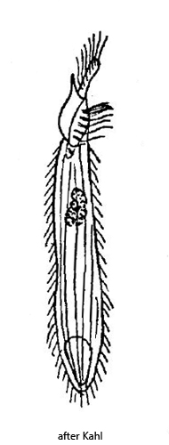
body slender cylindrical, slightly flattened, slightly tapered posterior end
often deformed by ingested food (separately ingested rhodobacteria)
anterior end flattened, tongue-shaped
length 100–110 µm
short adoral zone of 6 membranelles, reaching to the first sixth
cilia loosely arranged, slow swimming style
attaches thigmotactically to substrate
macronucleus short elliptic
contractile vacuole terminal
Metopus tenuis
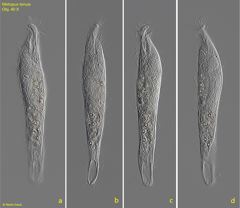
Kahl describes Metopus tenuis as “very rare”. Also I could detect Metopus tenuis only twice. The first finding was in February 2008 (s. fig. 1 a-b) and two further specimens in February 2022 (s. fig. 3 a-c) and April 2022 (s. fig. 1 a-d). All findings are from the Simmelried. In my other localities I could not detect Metopus tenuis so far.
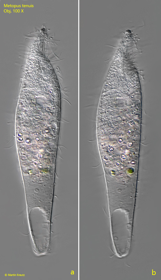
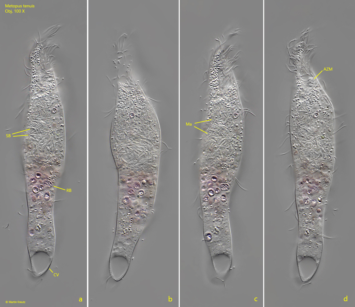
The first specimen with 170 µm length was clearly longer than the range of 100–110 µm given by Kahl. However, we do not know on how many observations Kahl’s length data are based. Therefore 170 µm may still be within the common range for this species. The second finding was only 102 µm long and the third 157 µm. In all specimens I could detect a high concentration of symbiotic bacteria around the macronucleus (s. fig. 3 a-d), which Kahl does not mention in his short description. I already made a similar observation in Metopus spinosus. Whether they are only temporarily present or only occur in certain populations/habitats cannot be said yet without having evaluated further findings.
In all specimens I found ingested rhodobacteria (s. fig. 3 a), which are phagocytosed individually according to Kahl’s description. I could not observe phagocytosis itself.
The adoral zone is very short (s. fig. 2 d) and hard to see in a slowly rotating specimen. However, characteristic of Metopus tenuis is the slender body and the elongated and flattened anterior end.
Fig. 1 a-d:Metopus tenuis. L = 157 µm. A freely swimming specimen found in April 2022. Obj. 40 X.
Fig. 2 a-b:Metopus tenuis. L = 170 µm. Ventral view of a freely swimming specimen found in February 2008. Obj. 100 X.
Fig. 3 a-d:Metopus tenuis. L = 102 µm. Ventral view of a freely swimming specimen found in February 2022. Note the symbiotic bacteria (SB) covering the macronucleus (Ma). AZM = adoral zone of membranelles, RB = ingested rhodobacteria. Obj. 100 X.