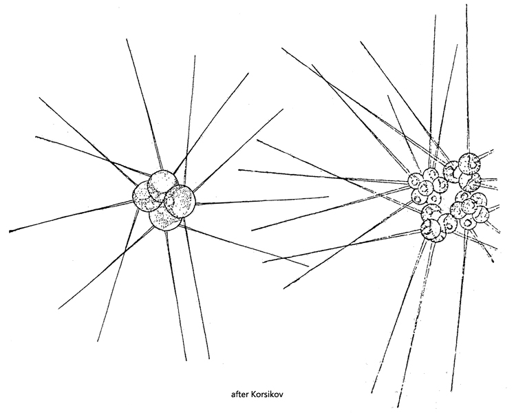
cells spherically, 3–13 µm in diameter, commonly 7–13 µm
cell wall smooth, bearing 2–8 hollow bristles
bristles 12–100 µm long, 0.5–2.5 µm wide, sometimes wider at base
one parietal chloroplast
one pyrenoid
nucleus central
planktonic lifestyle
Micractinium pusillum
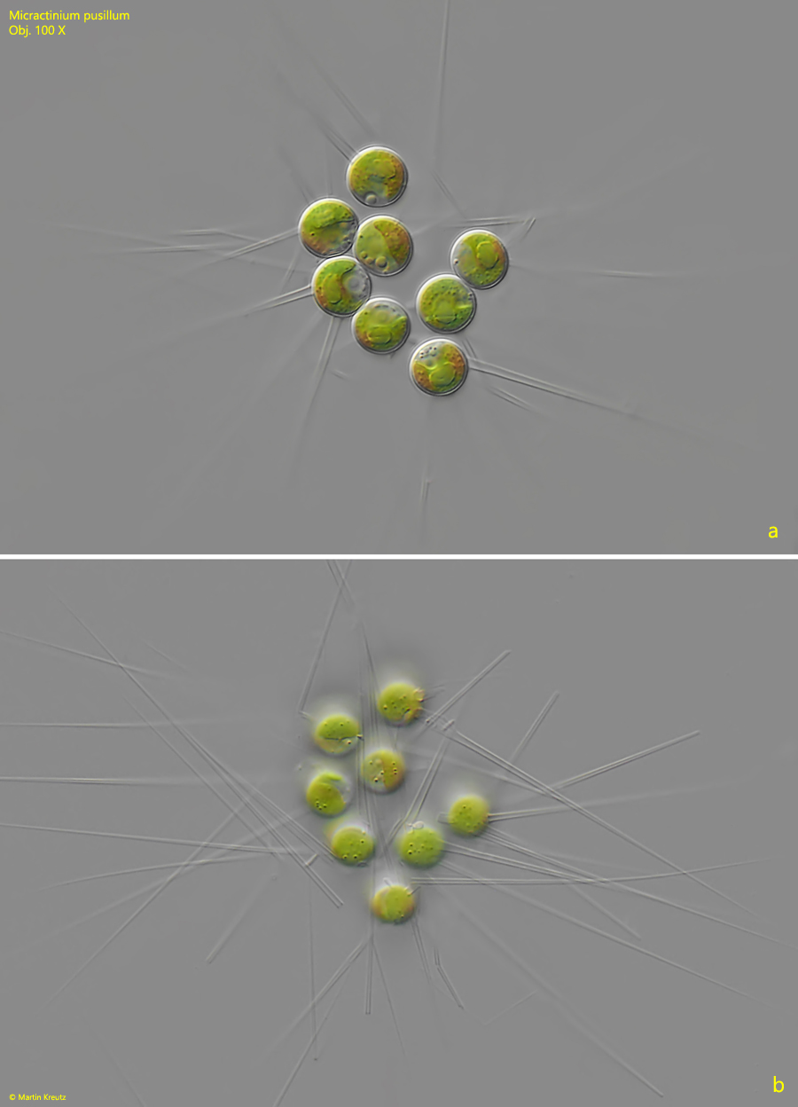
Micractinium pusillum is common in all of my sites, which allow for a planktonic lifestyle. This alga is especially common in the heavily eutrophic pond of waste disposal company Constance. Colonies are easily identified by their long, hollow bristles. These sit on the outer surface of the cell wall, but do not appear to be firmly attached to it. Slight mechanical force causes them to break away from it. In my populations I could also observe a basal broadening of the bristles. The cells are of the chlorella type, i.e. with a simple parietal chloroplast, a pyrenoid and the centrally located nucleus.
Fig. 1 a-b:Micractinium pusillum. D = 7–8 µm. Two focal planes of a slightly squashed colony. Obj. 100 X.
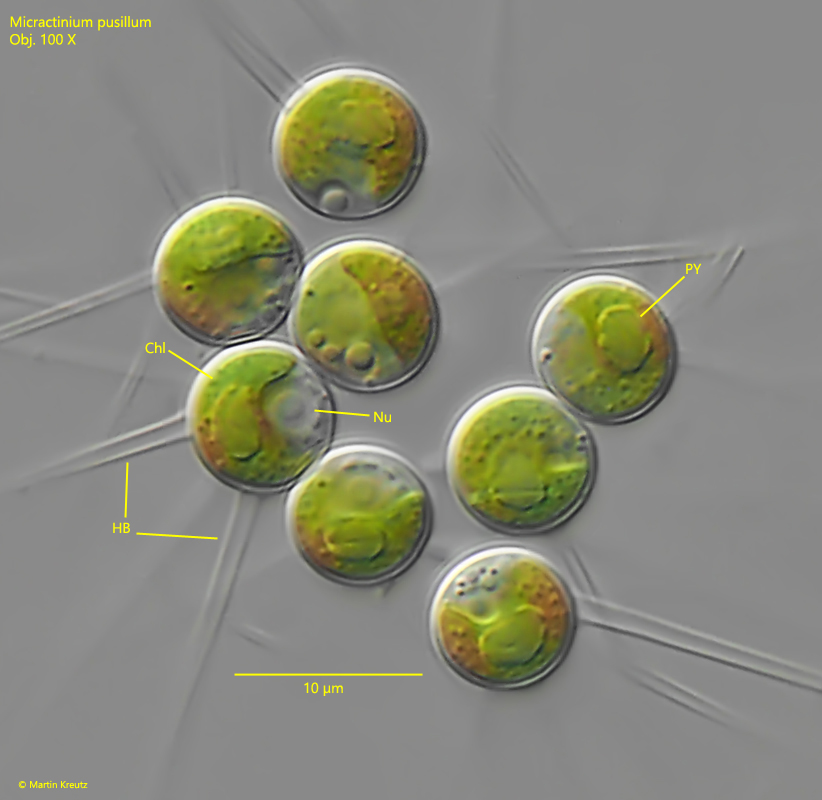
Fig. 2:Micractinium pusillum. D = 7–8 µm. The cells of the colony shown in fig. 1 in detail. Chl = chloroplast, HB = hollow bristles, Nu = nucleus, PY = pyrenoid covered with starch grains. Obj. 100 X.
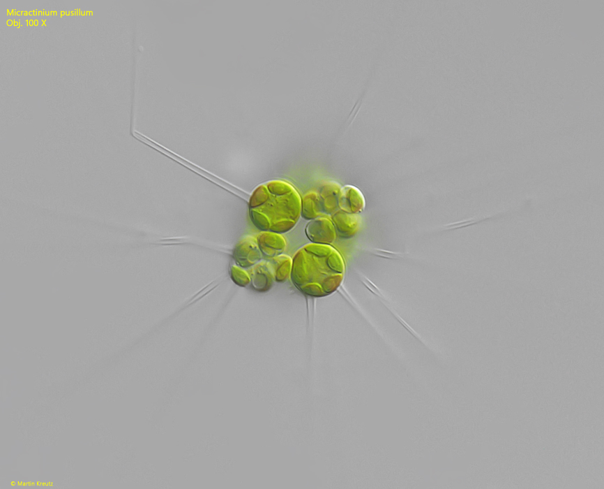
Fig. 3:Micractinium pusillum. A colony during the formation of autospores. Obj. 100 X.