Micrasterias rotata is one of the most common Micrasterias species and is very adaptable. Sometimes massive populations develop. Mainly it occurs in moorland areas and peat bogs. I found Micrasterias rotata in the nature reserve Schwemm near Walchsee in Austria.
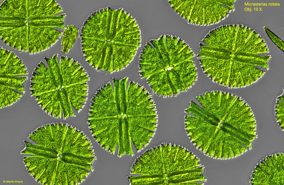
Fig. 1:Micrasterias rotata. Mass development in a sample from the nature reserve Schwemm. Obj. 10 X.
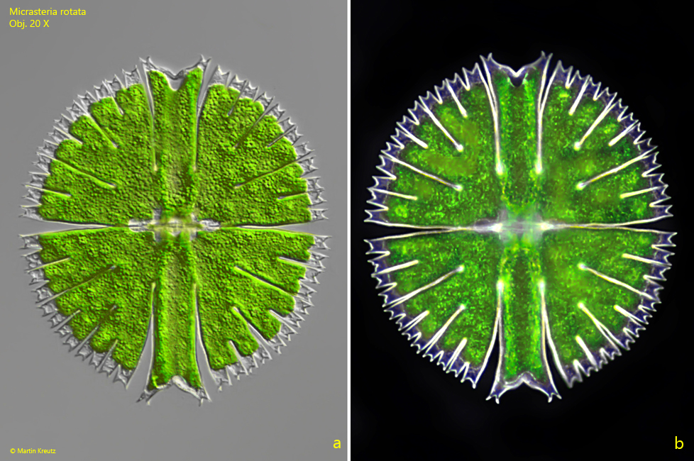
Fig. 2 a-b:Micrasterias rotata. L = 270 µm. a) in DIC, b) in dark field. Obj. 20 X.
Fig. 3 a-b:Micrasterias rotata. L = 244 µm. Two focal planes of a second specimen in brightfield illumination. Obj. 40 X.
Fig. 4 a-b:Micrasterias rotata. L = 244 µm. The specimen as shown in fig. 3 a-b in DIC. Obj. 40 X.
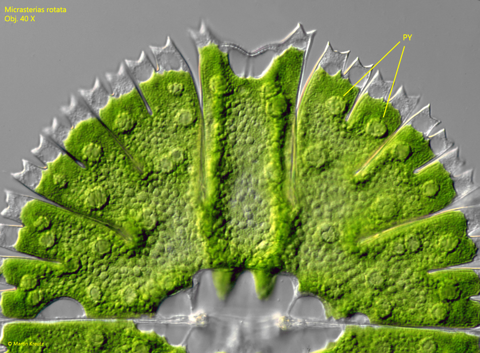
Fig. 5:Micrasterias rotata. Details of a semi-cell with scattered pyrenoids (PY). Obj. 40 X.
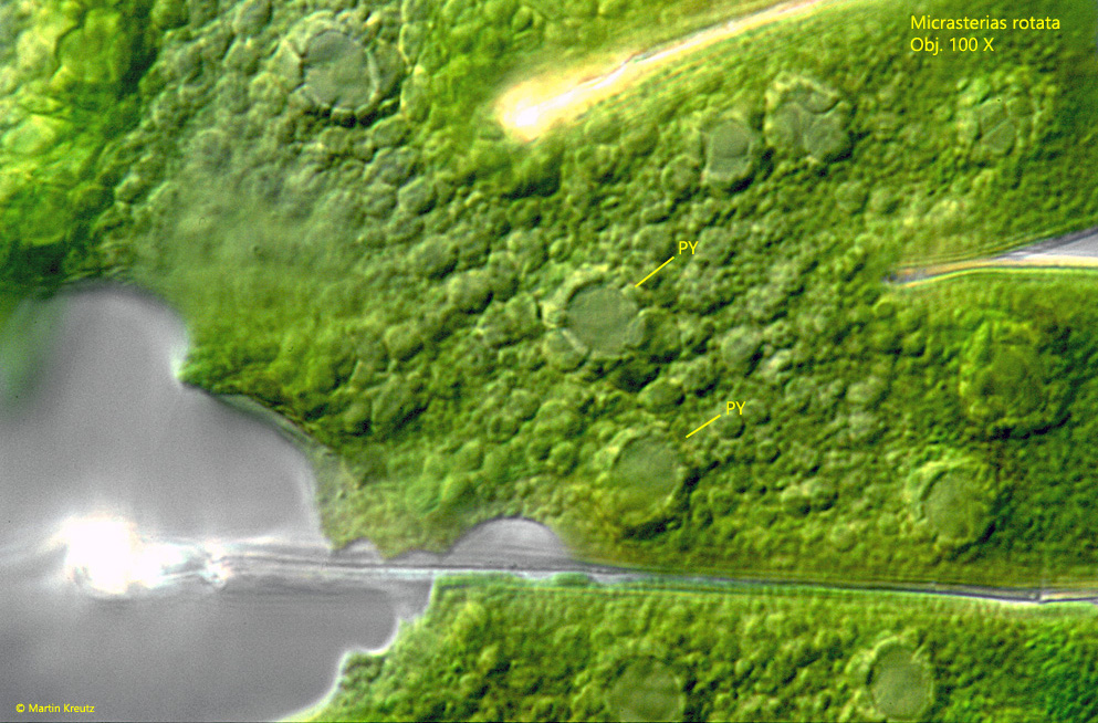
Fig. 6:Micrasterias rotata. The pyrenoids (PY) are covered with a layer of starch grains. Obj. 100 X.
Most desmids such as Micrasterias are surrounded by a gelatinous layer. The excretion of the jelly is often not only radially symmetrical as in Xanthidium, but also directional, e.g. at the cell ends. This results in a directed movement of the algae. Micrasterias rotata is covered by a gelatinous layer and also capable of such movement. The cells are often accumulates on the mud surface in dendrite-like structures.
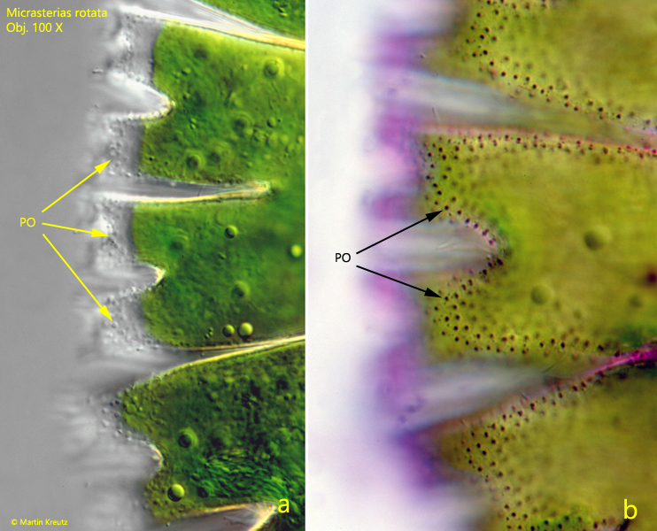
The gelatinous layer is secreted through pores, or more precisely by pore apparatuses. These provide a connection between cytoplasm and the outside of the cell wall. The jelly formed inside the cell is secreted through these pores. The pores can be recognized by light microscopy as dotting of the cell surface. These pore apparatuses can be easily stained by the basic dye crystal violet, because the jelly has acidic properties. By this approach, the pore apparatuses in the cell wall can be easily visualized.
Fig. 7:Micrasterias rotata. The pores on the cell surface in a unstained specimen (a) and after staining with crystal violet (b). Obj. 100 X.