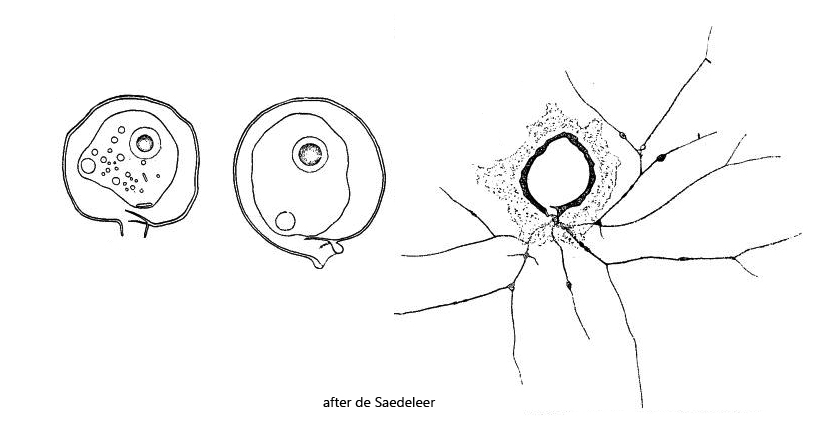
shell brownish with and thick layer of iron precipitation on older specimens
short neck, perpendicular or obliquely oriented to shell outline
neck with a septum, sometimes two septa
protoplast fills the shell only partially
nucleus central with a spherical nucleolus
contractile vacuole near neck
granuloreticulopodia very thin, anastomosing, arising from a peduncle
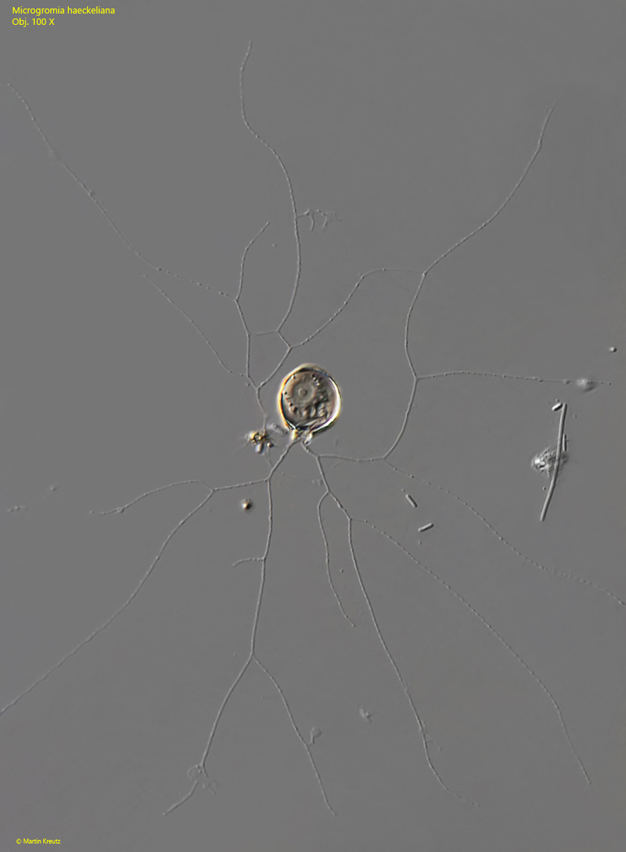
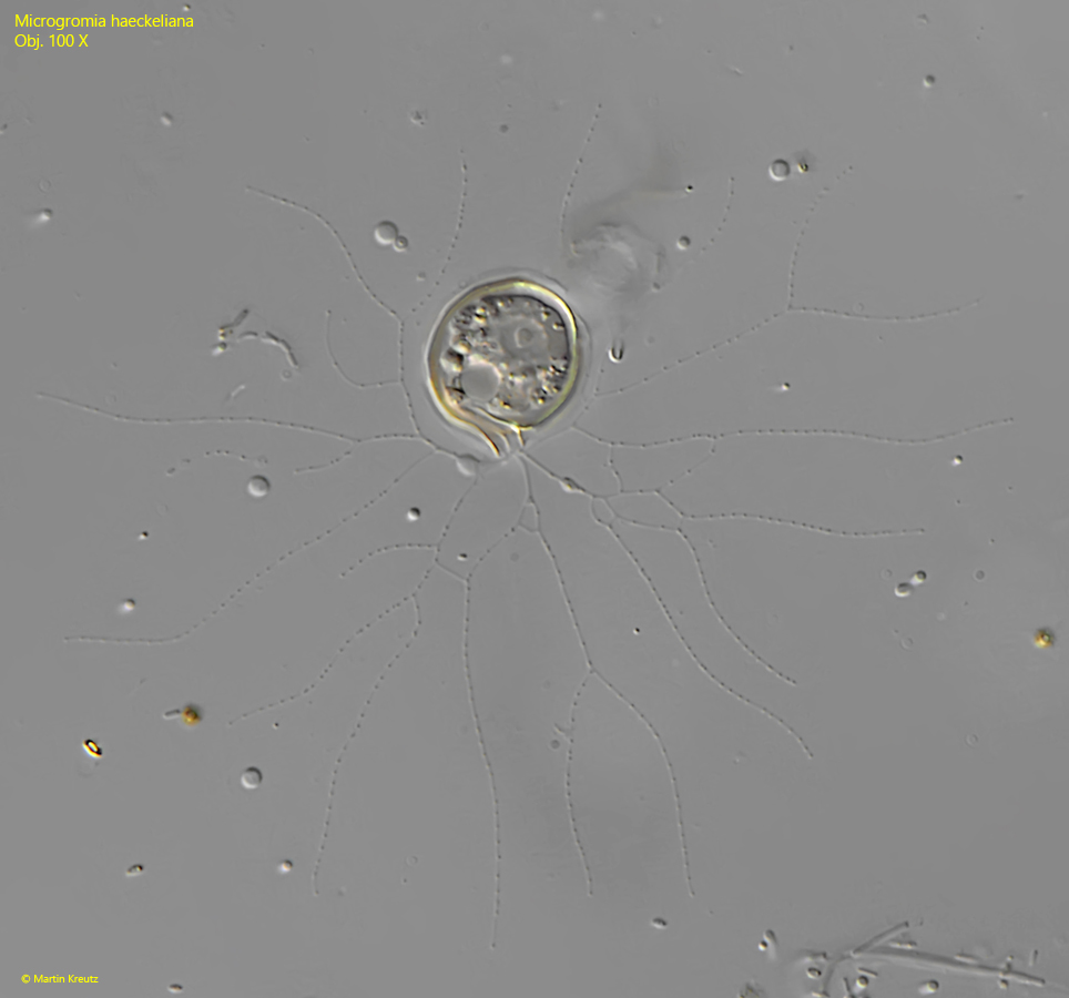
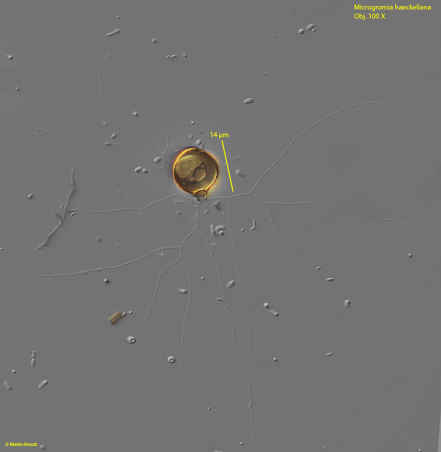
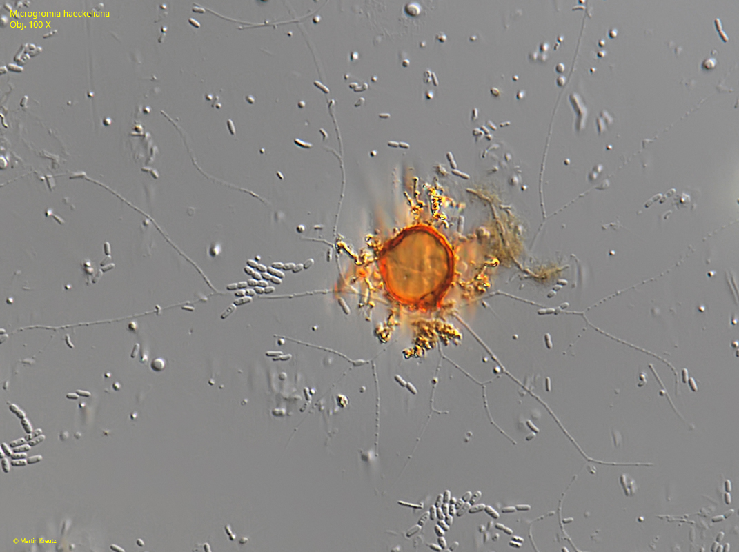
Microgromia haeckeliana
Microgromia haeckeliana is the most common species of the genus Microgromia in Simmelried. It occurs there frequently and regularly. As with almost all other small testate amoebae, the individuals are not found directly in the samples because they are often stuck in detritus flocs. However, if an aliquot of the sample is transferred into Petri dish and some floating coverslips are placed on the surface, Microgromia haeckeliana (and many other testate amoebae) will settle on it after only a few days.
Microgromia haeckeliana has a septum at the base of the neck, like all other Microgromia species. The short neck of Microgromia haeckeliana is oriented in a straight or oblique way to the shell and sometimes it is thickened at the distal margin (s. figs. 4 and 5). Although de Sadeleer drew and described specimens with two septa (s. drawings above), I could only observe specimens with one septum in my population. Another characteristic peculiarity of Microgromia haeckeliana is the accumulation of iron precipitates on the shell (iron oxide and iron hydroxide). This causes the shells to turn more and more orange or brown with age (s. fig. 7). In extreme cases the layer of iron precipitates can be so thick that the neck disappears in it (s. fig. 8).
Fig. 1:Microgromia haeckeliana. L = 14 µm. A fully expanded specimen. Note the widely branched granuloreticulopodia. Obj. 100 X.
Fig. 2:Microgromia haeckeliana. L = 18 µm. A second fully expanded specimen. Obj. 100 X.
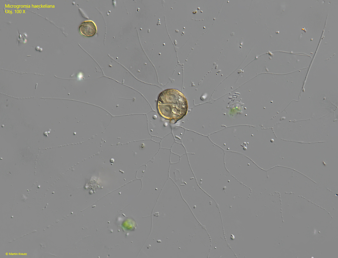
Fig. 3:Microgromia haeckeliana. A third fully expanded specimen. Obj. 100 X.
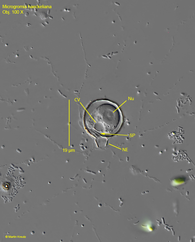
Fig. 4:Microgromia haeckeliana. A specimen with a clearly visible septum (SE). Note that the protoplast fills the shell only partially. CV = contractile vacuole, NE = neck, Nu = nucleus. Obj. 100 X.
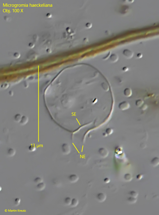
Fig. 5:Microgromia haeckeliana. An empty shell with a clearly visible septum (SE. NE = neck. Obj. 100 X.
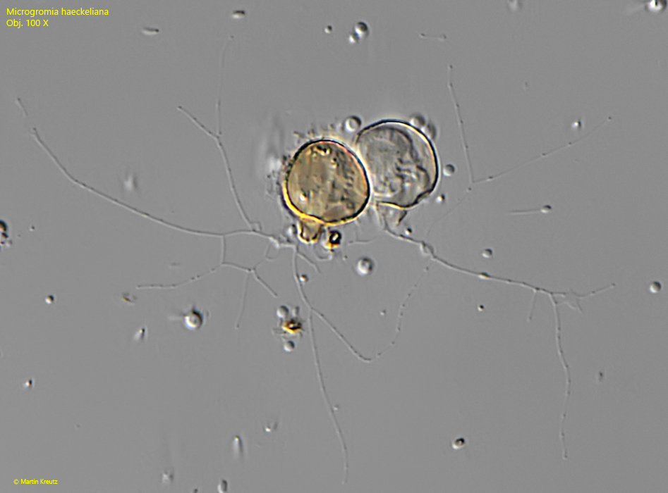
Fig. 6:Microgromia haeckeliana. L = 16 µm. A shell with two specimens (two nuclei) after cell division. Obj. 100 X.
Fig. 7:Microgromia haeckeliana. L = 12 µm. Two specimens with different degrees of coloration by iron precipitations. Obj. 100 X.
Fig. 8:Microgromia haeckeliana. L = 14 µm. A specimen what is strongly covered by iron precipitations. Obj. 100 X.