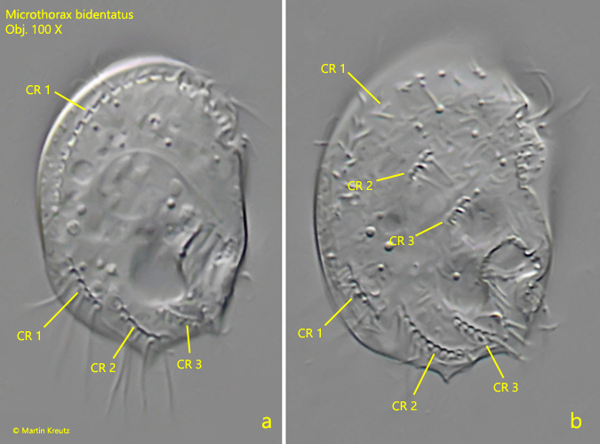
one ciliary row on right side parallel to the convex dorsal margin
two short ciliary rows in the center of the right side
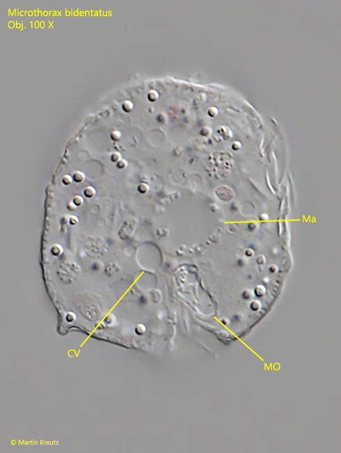
spherical macronucleus in center of cell
contractile vacuole above mouth opening
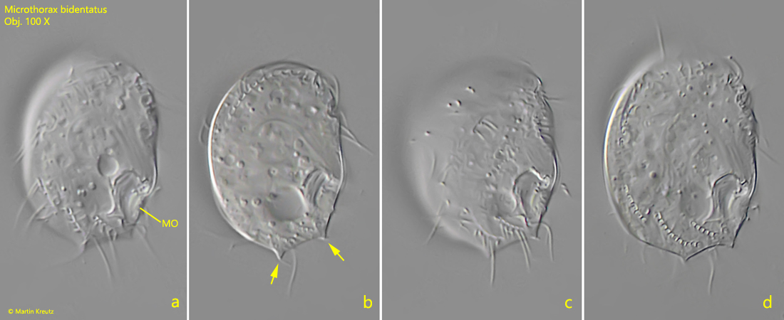
oral apparatus at posterior end of cell
Microthorax bidentatus
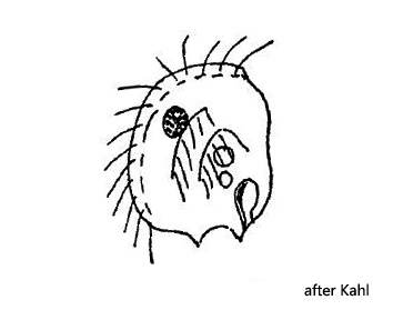
So far I have found Microthorax bidentatus exclusively in the Simmelried. However, I have found only a few specimen over the years. The last time I found the species in November 2019 and in December 2022. I may have missed it earlier, as the two characteristic teeth at the posterior end are only visible at higher magnifications. In the literature I have found only the very short description by Kahl, who also gives only a single drawing from the right side (s. drawing above). The individuals in my population differ partly from the description of Kahl. He gives a size of 16 µm, while my individuals were 28 – 31 µm long. The ciliation of the right side Kahl only drew without giving any further description on it. On his drawing one can recognize a continuous dorsal row of cilia, which runs parallel to the dorsal margin. He also draws two other short rows, approximately in the middle of the cell. According to my observations, the dorsal row is interrupted in the middle and the two rows beginning approximately in the middle of the cell find a continuation in the posterior third after an interruption (s. fig. 2 a-b).
Fig. 1 a-d:Microthorax bidentatus. L = 31 µm. Lateral view of the right side of a freely swimming specimen. Note the distinct two teeth at the posterior end (arrows). MO = mouth opening. Obj. 100 X.
Fig. 2 a-b:Microthorax bidentatus. L = 31 µm. The ciliation of the right side. There are three ciliary rows (CR 1 – CR 3). The ciliary row CR 1 runs parallel to the convex dorsal margin and is interrupted in the middle. The ciliary row CR 2 starts in the anterior third of the cell and is interrupted in the mid-body region. The ciliary row CR 3 starts in the mid-body region and is interrupted in the posterior third of the cell. Obj. 100 X.
Fig. 3:Microthorax bidentatus. L = 31 µm. A strongly squashed specimen for visualisation of the spherical macronucleus (Ma) in the center of the cell. CV = contractile vacuole, MO = mouth opening. Obj. 100 X.