shape semicircular, ventral margin almost straight, dorsal convex
laterally flattened
length about 40 µm
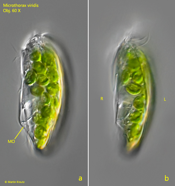
ventrally 2 preoral furrows with rows of cilia
right side flat with 3 rows of cilia interrupted in middle
left side flat with an apical indentation and 5 pores each with one cilium
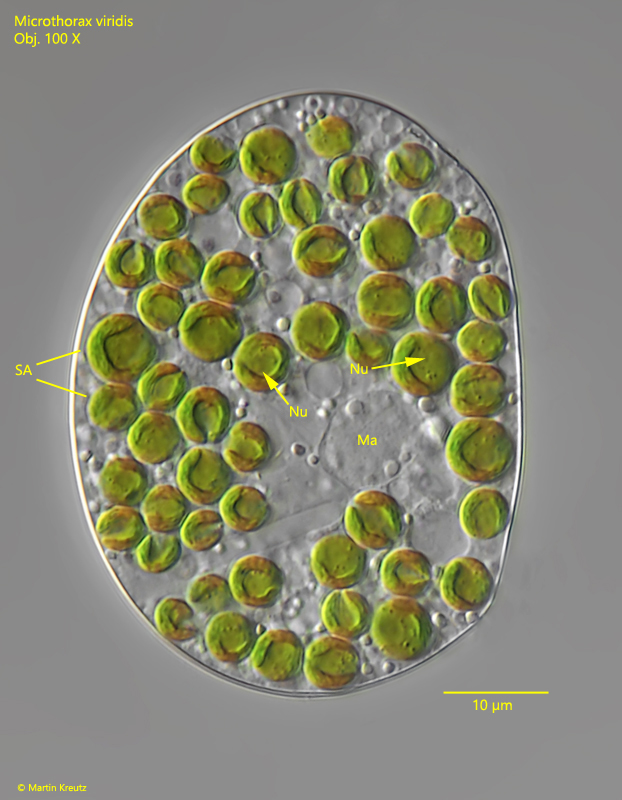
cytoplasm filled with 40–60 symbiotic algae
macronucleus spherical in mid-body
contractile vacuole almost mid-body
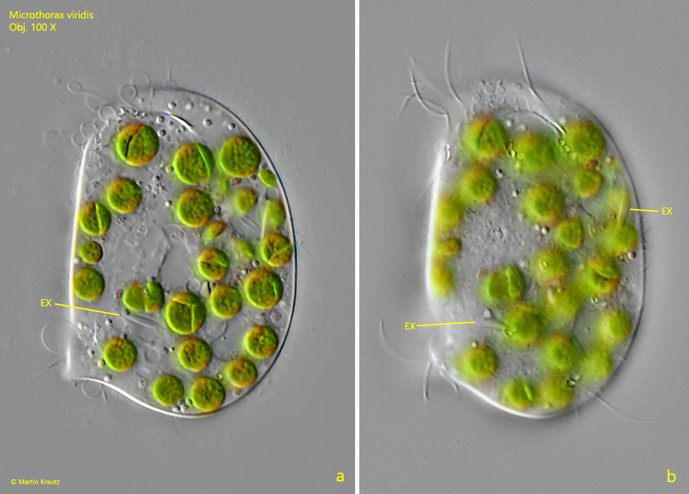
some spindle-shaped extrusomes below the pellicle
oral apparatus at posterior end of cell shifted to right side
Microthorax viridis
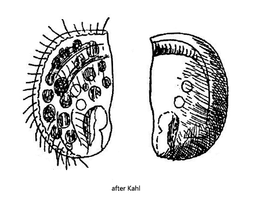
Microthorax viridis is one of the most common ciliates in the mud from Simmelried. The species is present in almost every sample. Despite its small dimensions of about 40 µm, Microthorax viridis can be recognized by its green color and characteristic shape even at low magnifications. Kahl gives only a very short description of Microthorax viridis and only a rough drawing of the left side (s. drawing above). On the right side (fg. 1 a-b and 2 a-b) 3 rows of cilia are visible, which are all interrupted in the middle of the body (s. fig. 1a). To my knowledge the left side has not been examined and described yet. There I could observe and document 5 pores with one cilium each (s. fig. 3 a-b). As a consequence two pores are in the anterior third and three pores in the posterior half. The number of symbiotic algae I could determine to be 40–60. Mostly there were about 50 symbiontic algae. The symbiotic algae have a diameter of 5–7 µm and have their own nucleus (s. fig. 4). They seem to be of Chlorella type. I could detect some spindle-shaped extrusomes with a length of 6 µm below the pellicle (s. fig. 5 a-b). However, the number of extrusomes per cell was < 10.
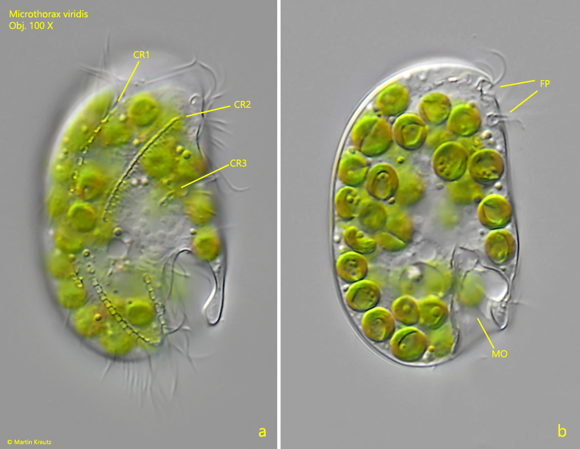
Fig. 1 a-b: Microthorax viridis. L = 38 µm. Two focal planes of the right side. Note the three interrupted rows of cilia (CR1 – CR3) and the two ventral furrows with preoral rows of cilia (FP). MO = mouth opening. Obj. 100 X.
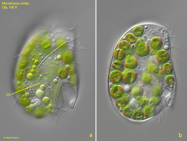
Fig. 2 a-b: Microthorax viridis. L = 35 µm. Two focal planes of the right side of a second specimen. CV = contractile vacuole. Obj. 100 X.
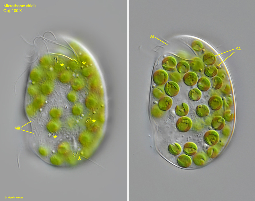
Fig. 3 a-b: Microthorax viridis. L = 43 µm. Two focal planes of the left side. Note the five pores each with one cilium (labelled with *) and the apical indentation (AI). Mit = mitochondria beneath the pellicle, SA = symbiotic algae. Obj. 100 X.
Fig. 4: Microthorax viridis. A strongly squashed specimen from the right side. This specimen contains 53 symbiotic algae (SA) of the Chlorella type with a diameter of 5–7 µm. The nuclei of the symbiotic algae are visible (Nu). Ma = macronucleus. Obj. 100 X.
Fig. 5 a-b: Microthorax viridis. L = 43 µm. Two focal planes of a slightly squashed specimen from the left side. Below the pellicle some spindle-shaped extrusomes (EX) are visible with a length of 6 µm. Obj. 100 X.
Fig. 6 a-b: Microthorax viridis. L = 43 µm. Two focal planes of a freely swimming specimen from ventral demonstrates the laterally flattened shaped of the cell. MO = mouth opening. Obj. 100 X.