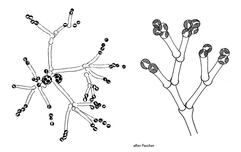
cells attached to distal end of dichotomously branched mucilaginous stalks
1–2 parietal chloroplasts, pale yellow, without pyrenoid
branched colonies often attached to filamentous algae
swarmer with two flagella and two contractile vacuoles
Mischococcus confervicola
I rarely find Mischococcus confervicola in my samples. The colonies are usually found between floating plants or in algae tufts. The characteristic growth form makes this xanthophyte easy to identify, although the cells are very small. In my population mostly between 6–8 µm. The cells are located at the ends of mucilaginous stalks, which are excreted by the cells. These mucilaginous stalks branch dichotomously at different angles, creating the characteristic appearance of the colonies.
Fig. 1:Mischococcus confervicola. D = 210 µm (of colony). A slightly squashed colony. Obj. 40 X.
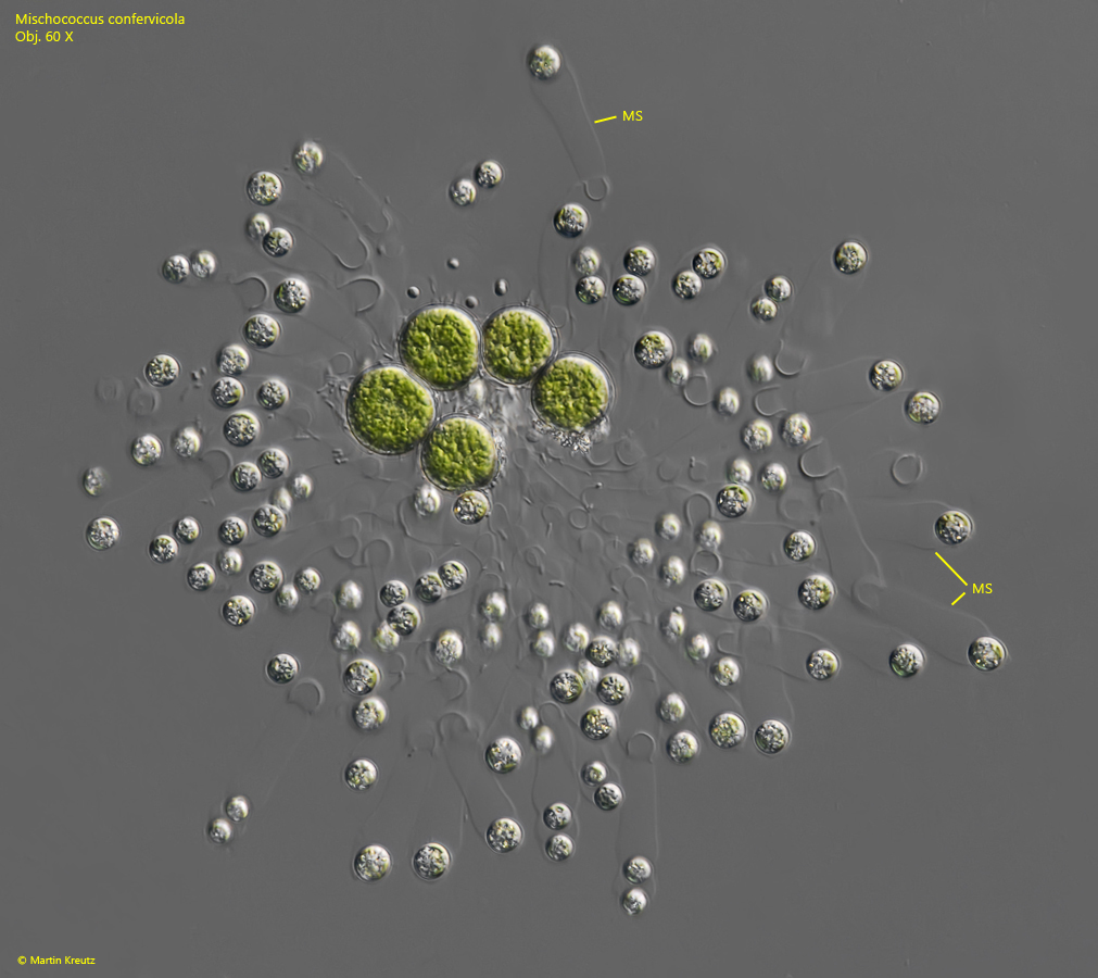
Fig. 2:Mischococcus confervicola. D = 220 µm (of colony). A second, slightly squashed colony growing of a cluster of 5 cells of chlorophytes. Note the mucilaginous stalks (MS) of the cells. Obj. 60 X.
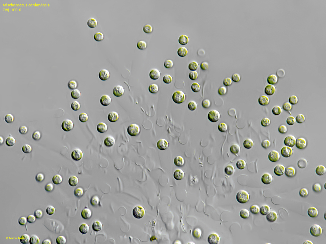
Fig. 3:Mischococcus confervicola. The spherical cells in a strongly squashed colony. Obj. 100 X.
Fig. 4 a-b:Mischococcus confervicola. D = 5.7 – 6.8 µm (of cells). The cells in detail. Note the very small nucleus (Nu) in the center of the cell. Chl = chloroplasts. Obj. 100 X.