single oval macronucleus and one spherical micronucleus
Mylestoma anatinum
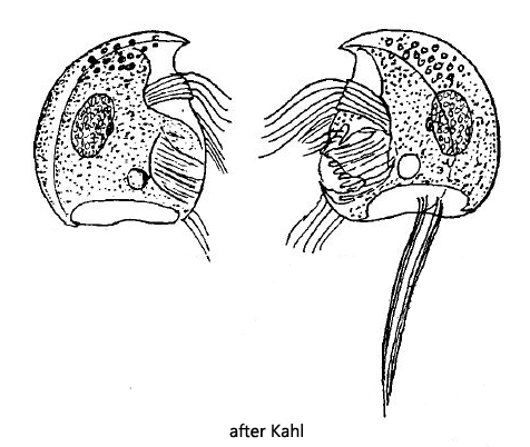
Mylestoma anatinum ist common in the mud of Simmelried. The species can be regognized by the lack of dorsal spine and a large, posterior indentation (s. fig. 4a). Two cirri are located at the posterior end of the left side (s. fig. 1a-c and fig. 2 a-b). According to Kahl Mylestoma anatinum reaches a length of up to 25 µm. In my population the specimens reached a length of 21–27 µm what is within the common variation.
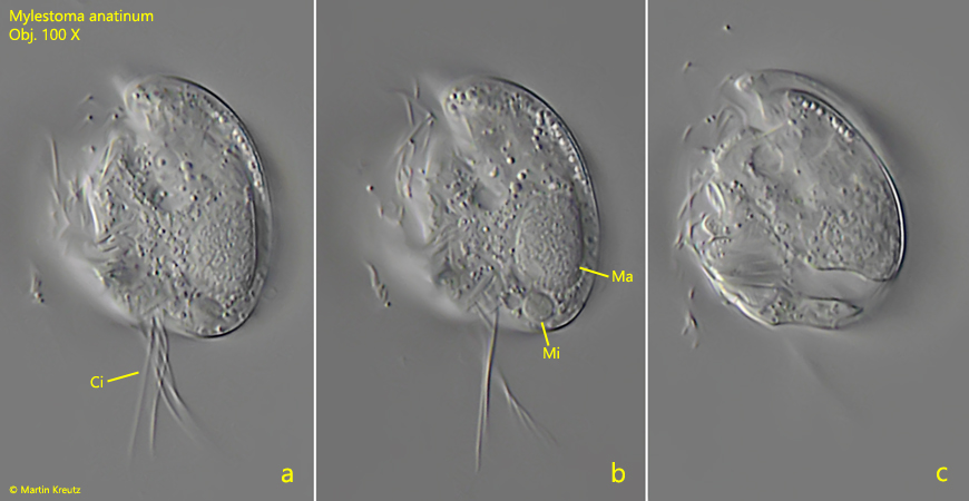
Fig. 1 a-c: Mylestoma anatinum. L = 25 µm. Three focal planes of a freely swimming specimen from the left side. Ci = posterior cirri, Ma = macronucleus, Mi = micronucleus. Obj. 100 X.
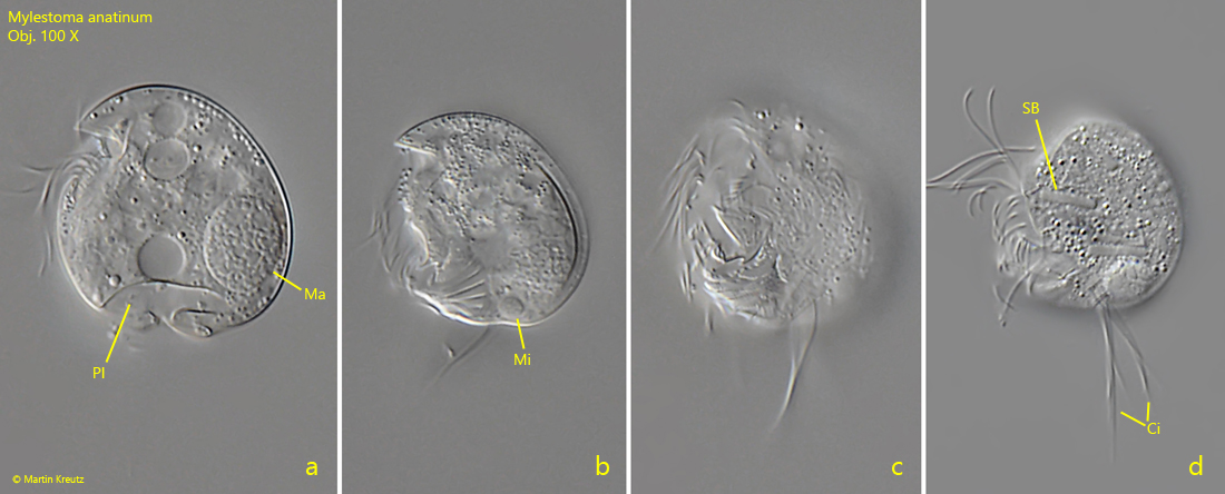
Fig. 2 a-d: Mylestoma anatinum. L = 22 µm. Four focal planes of a slightly squashed second specimen from the left side. Ci = posterior cirri, Ma = macronucleus, Mi = micronucleus, PI = posterior indentation, SB = symbiotic bacteria. Obj. 100 X.
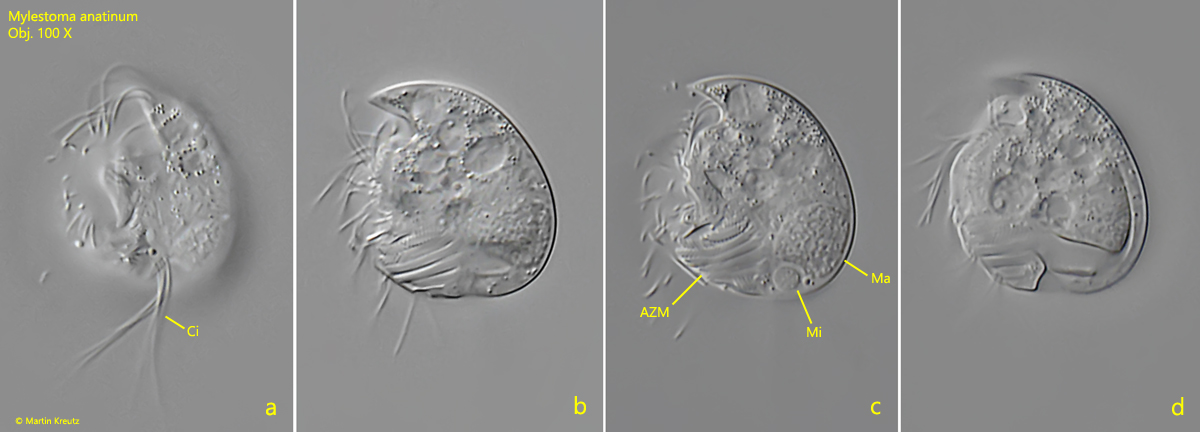
Fig. 3 a-d: Mylestoma anatinum. L = 21 µm. Four focal planes of a third specimen from the left side. AZM = adorale zone of membranelles, Ci = posterior cirri, Ma = macronucleus, Mi = micronucleus. Obj. 100 X.
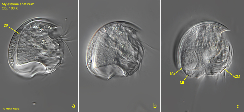
Fig. 4 a-c: Mylestoma anatinum. L = 27 µm. Three focal planes of a slightly squashed specimen from the right side. AZM = adorale zone of membranelles, DR = dorsal ridge, Ma = macronucleus, Mi = micronucleus. Obj. 100 X.
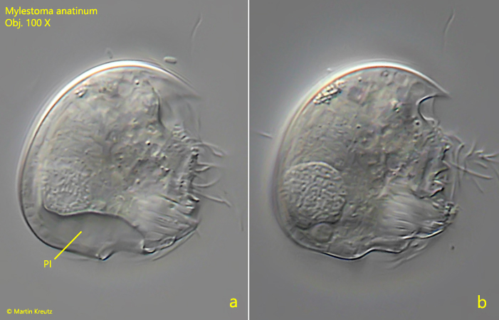
Fig. 5 a-b: Mylestoma anatinum. L = 26 µm. Two focal planes of a second specimen from the right side. PI = posterior indentation. Obj. 100 X.
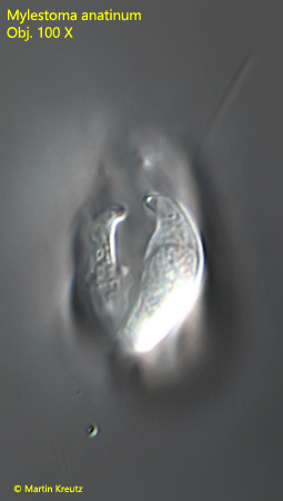
Fig. 6: Mylestoma anatinum. Posterior view of a freely swimming specimen. Obj. 100 X.