two long posterior cirri behind oral apparatus on left side
without spines
single macronucleus and micronucleus
Mylestoma dicoideum
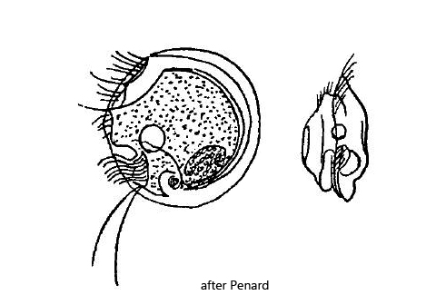
There are only a few observations of the odontostomatid ciliate Mylestoma discoideum, all of which were made by Penard and Kahl. Kahl found only one specimen. I also found only three specimens in August 2018 and in September 2019 in the Simmelried. After 2019 I found no further specimens and in my other sampling sites Mylestoma discoideum is not present.
The characteristics of the specimens agreed to a large extent with the drawings of Penard (s. drawings above). However, my specimens were with 18–21 µm much smaller than those of Penard, who gives 35–38 µm as size. On the other hand the specimen found by Kahl was also only 27 µm long.
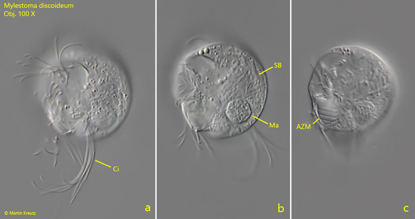
An important characteristic of Mylestoma discoideum is the almost circular shape of the body without any spines. The round shape of the dorsal side extends almost to the oral apparatus. On the posterior end of the left side there are long cirri. Penard draw only 2 long cirri (s. drawing above) and Kahl mentions that he may have missed them. I could clearly recognize a tuft of long cirri (s. fig. 1 a), even if they are not as close to the oral apparatus as Penard drew them. Since there are so few observations, it remains unclear for now whether this is within the variance of the species. Kahl also mentions that the ventral line appears angular. I can confirm this observation (s. fig. 1 c). In the cytoplasm, I found many symbiotic bacteria (s. fig. 1 b) explicitly mentioned by Kahl, although those are obviously present in all odontostomatid ciliates.
Fig. 1 a-c: Mylestoma discoideum. L = 21 µm. Three focal planes of the left side of a slightly squashed specimen. AZM = adoral zone of membranelles, CI = posterior cirri on left side, Ma = macronucleus, SB = symbiotic bacteria. Obj. 100 X.
Fig. 2 a-c:Mylestoma discoideum. L = 21 µm. Three focal planes from the right side of a slightly squashed specimen. In fig. 5 c the focal plane is on the left side seen from the right side. Ma = two macronuclei or a macronucleus with adjacent micronucleus, PC =long posterior cilia of the left side RR = ridge of the right side. Obj. 100 X.
Fig. 3 a-e:Mylestoma discoideum L = 18 µm. Different focal planes of a third specimen from the left side. Ma = macronucleus, Mi = micronucleus, PC = long posterior cilia. Obj. 100 X.