Mylestoma pusillum (Kahl, 1931)

Most likely ID: n.a.


Synonym: n.a.


Sampling location: Simmelried


Phylogenetic tree: Mylestoma pusillum


Diagnosis:

  • body oval, laterally flattened
  • length 14 – 20 µm
  • pellicle armour-like
  • no posterior spines
  • no frontal tooth
  • no indentations at posterior end
  • C-shaped longitudinal ridge on right side
  • one long cirrus (2 or 3 Cilia) at posterior end of left side
  • two long dorsal cilia
  • large peristom with prominent adoral zone of membranelles
  • single macronucleus
Mylestoma-pusillum
Mylestoma pusillum

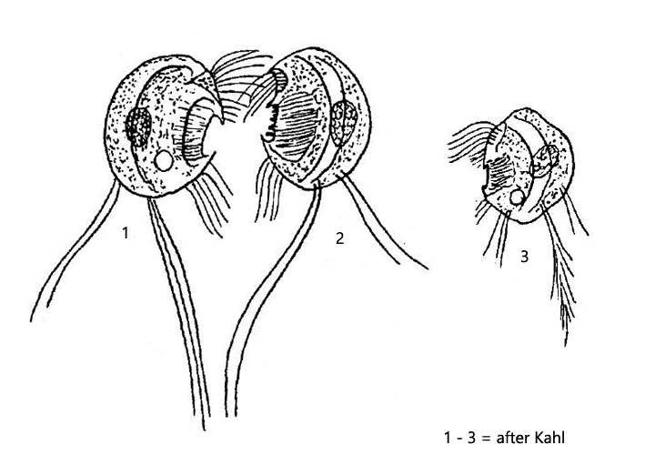
The very small odontostomatid ciliate Mylestoma pusillum has been studied in more detail only by Kahl, who describes the species as “very rare”. One must assume that his description is based on the examination of a few specimens. I found this species exclusively in the mud of Simmelried. It appears there sporadically at longer intervals, but then in large numbers. I found populations so far in October 2008, June 2018, August 2018 and most recently in August 2019.

In my opinion, the drawings of Mylestoma pusillum by Kahl (s. drawings above) are quite different from the real shape, as he drew it very oval. However, in living specimens the posterior end appears truncated. But his exact description of the other characteristics allows a definite identification. The species has a C-shaped ridge on the right side (s. drawing 1, figs. 1a and 3a). On the left side there is a very prominent groove (s. drawing 2, figs. 2a and 4a) and 2-3 cilia at the posterior end, which can reach twice the body length (see fig. 4b). At the dorsal margin there are more, shorter cilia. According to my observations there seem to be 2 – 4 cilia. The oral openung with the adoral membranelles is very large in relation to the size of the ciliate (s. figs. 2c and 3c).


The species can be recognized already at smaller magnifications (< Obj. 40) by the very long cilia at the posterior end. Mylestoma pusillum has a trembling swimming style and drags these long cilia behind.
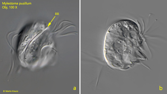
Mylestoma-pusillum

Fig. 1 a-b: Mylestoma pusillum. L = 15 µm. Two focal planes from the right side of a slightly squashed specimen. Note the longitudinal ridge on the right side (RR). Obj. 100 X.
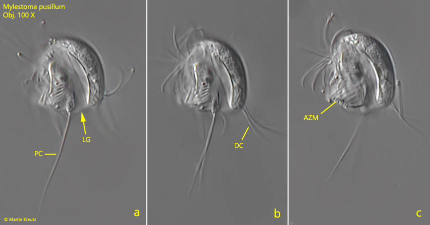
Mylestoma-pusillum

Fig. 2 a-c: Mylestoma pusillum. L = 15 µm. Three focal planes from the left side of a slightly squashed specimen. Note the longitudinal groove (LG), the long posterior cilia (PC) and two long dorsal cilia (DC). AZM = adoral zone of membranelles. Obj. 100 X.
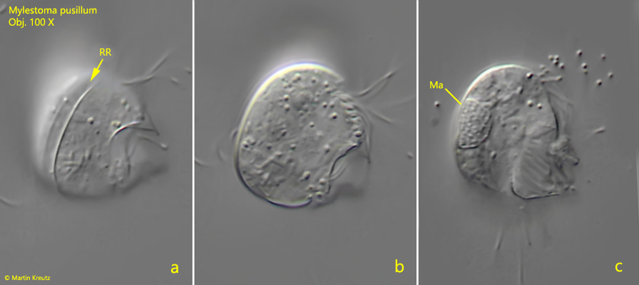
Mylestoma-pusillum

Fig. 3 a-c: Mylestoma pusillum. L = 16 µm. Three focal planes from the right side of a second specimen. RR = longitudinal ridge, Ma = macronucleus. Obj. 100 X.
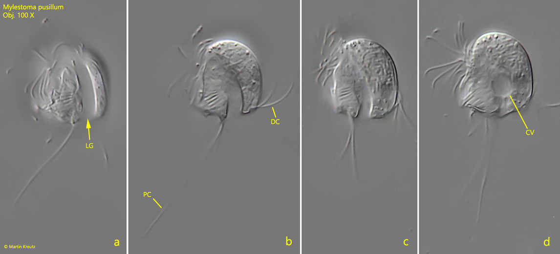
Mylestoma-pusillum

Fig. 4 a-d: Mylestoma pusillum. L = 15 µm. Four focal planes from the left side of a second specimen. CV = contractile vacuole, DC = dorsal cilia, LG = longitudinal groove, PC = posterior cilia. Obj. 100 X.

Kahl has made an additional drawing of Mylestoma pusillum (s. drawing 3) which is based on the finding of a single specimen. He reproduces here the left side, which he also draws with a long furrow. However, he describes and draws the cilia at the posterior end as “split like feathers”. He has also mistaken this form for Mylestoma pusillum, but also writes that this “needs further verification”. I could also find this form in Simmelried (in August 2018) however also only two specimens (s. figs. 5 a-c and 6 a-e). Both specimens were on the left side and I focused through them from the right side. In the first specimen, I documented the right side where there is a longitudinal ridge (s. fig. 5a). There is a deep incision of the dorsal margin. In any case, the body shape is oval as Kahl draws it. At the posterior end of the left side of this first specimen I could see the feathery tuft of cilia as Kahl drew it (s. fig. 5c). However, these were of maximum body length and there were also more than 3 cilia. I could not make out any other cilia on the left side. The specimen seemed to have two macronuclei (s. fig. 5b). However, after finding the second specimen with a very large micronucleus (s. fig. 6 d), it may be a macro- and micronucleus. The second specimen (s. fig. 6 a-e) was freely swimming due to the finding situation, which reduced the resolution. I focused from the right side to the left side. This specimen also possessed a feathery tuft of cilia on this side (s. fig. 6a). The body shape was nearly circular with a diameter of 18 µm. As with the first specimen, I could not see a clearly defined groove on the left side as described for Mylestoma pusillum. This may be due to the fact that I had to focus through the ciliate. Dorsal cilia could not be seen on either specimen.

From my previous observations on Mylestoma pusillum the shape seems to be very constant and corresponds to the images in Fig. 1-4. The oval shaped form, which are shown in figs.  5 and 6, seem to me to deviate so much that I do not believe that here is a shape variant of Mylestoma pusillum. The characteristics documented so far differ too much for that. However, to decide whether a separate species is present, a closer examination of the left side is necessary and examination of the ciliature is pending.

Mylestoma-cf-pusillum

Fig. 5 a-c: Mylestoma cf. pusillum. L = 21 µm. Three focal planes from the right side of a slightly squashed specimen. The fig. 5c is mirrored to give an impression of the left side. Ma = macronuclei (or a macronucleus with adjacent micronucleus), PC =long posterior cilia left side. Obj. 100 X.

Mylestoma-cf-pusillum

Fig. 6 a-e: Mylestoma cf. pusillum. L = 18 µm. Five focal planes of the left side through the ciliate. All images are mirrored to give an impression of the left side. Ma = macronucleus, Mi = micronucleus, PC = long posterior cilia. Obj. 100 X.