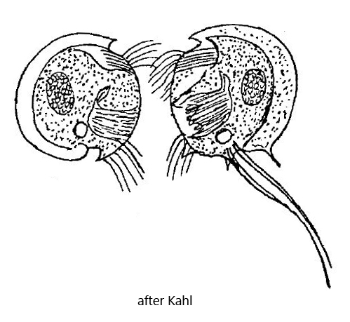
Kahl found Mylestoma uncinatum only sporadically. However, it is more common in deeper mud layers in my sampling site Purren pond. Possibly it is also present in my other sampling sites and I have overlooked the species because of its small size. In general, this species does not seem to have been observed frequently. The last sighting and description after Penard seems to be from Kahl.
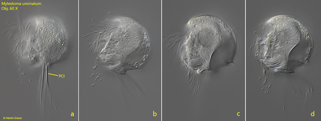
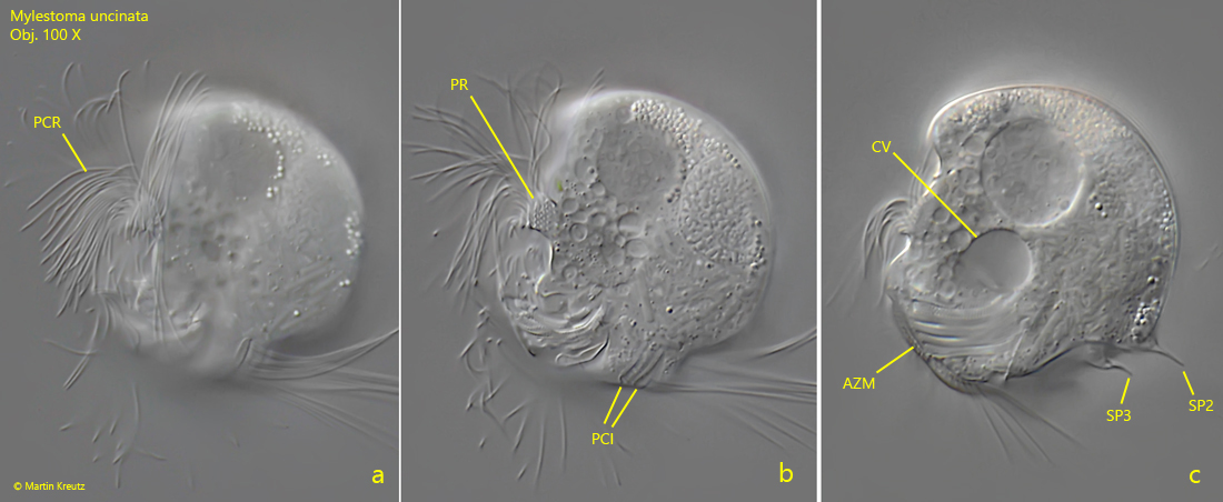
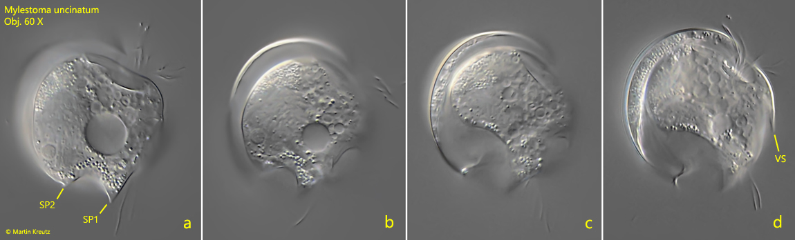
Freely swimming specimens of Mylestoma uncinatum rotate rapidly around their axis and appears lenticular. Identification is mainly based on the two prominent tufts of cilia arising from the posterior end on the left side, which are called cirri by Kahl (s. fig. 1a). This odontostomatid ciliate has only inconspicuous, short spines. There is a ventral spine (s. fig. 3d) that is difficult to see and is described by Kahl as “crenate.” There should be two spines at the posterior end according to Kahl (s. drawing above). According to my observations there are two spines (SP2 and SP3, s. fig. 2c) pointing dorsally. One of them seems to be on the left side and the other on the right side. In squashed specimens, however, it may appear that they both arise on the left side (s. fig. 2c). There is also a triangular tooth-shaped spine (SP1, s. figs. 3a and 4 b), which is on the right side. The perizonal stripe is short with quite long cilia arising from it. In the plasma I could observe large symbiotic bacteria (s. fig. 4a and fig. 5). They are rod-shaped and 3.8 – 6.2 µm long. The nucleus is simple and oval as drawn by Kahl. I could not identify the micronucleus.
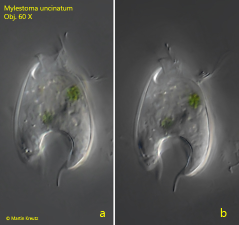
Fig. 1 a-d:Mylestoma uncinatum. L = 36 µm. Four focal planes of the left side of a freely swimming specimen. Note the prominent two cirri (PCI). Obj. 60 X.
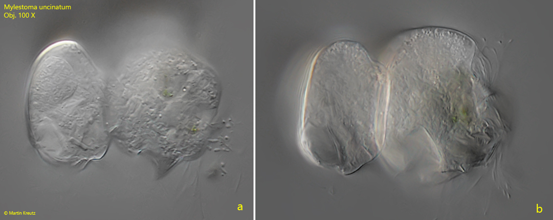
Fig. 2 a-c:Mylestoma uncinatum. L = 36 µm. Three focal planes of the left side of a slightly squashed specimen. Note the two posterior spines SP2 and SP3 (fig. 1c) pointing dorsally. AZM = adoral zone of membranelles, CV = contractile vacuole, PC = perizonal row, PCI = posterior cirri on left side, PCR = perizonal cilia row, PR = perizonal stripe. Obj. 100 X.
Fig. 3 a-d:Mylestoma uncinatum. L = 36 µm. Four focal planes of the right side of a freely swimming specimen. Note the ventral spine (VS). Obj. 60 X.
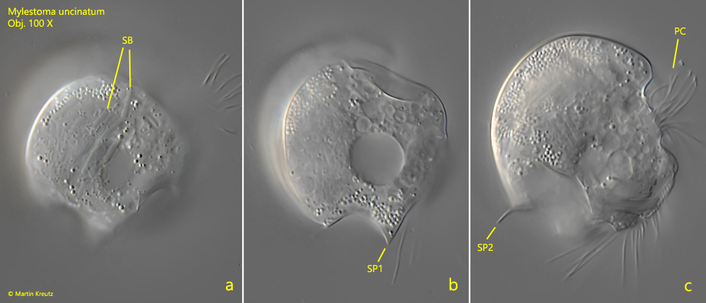
Fig. 4 a-c:Mylestoma uncinatum. L = 36 µm. Three focal planes of the right side of a slightly squashed specimen. Note the tooth-shaped spine 1 (SP1). PC = perizonal cilia, SB = symbiotic bacteria, SP2= spine pointing dorsally. Obj. 100 X.
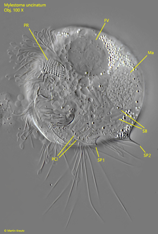
Fig. 5:Mylestoma uncinatum. L = 36 µm. A strongly squashed specimen from the left side. FV = food vacuole, Ma = macronucleus, PCI = posterior cirri, PR = perizonal stripe, SB = symbiotic bacteria, SP1, SP2 = posterior spines. Obj. 100 X.
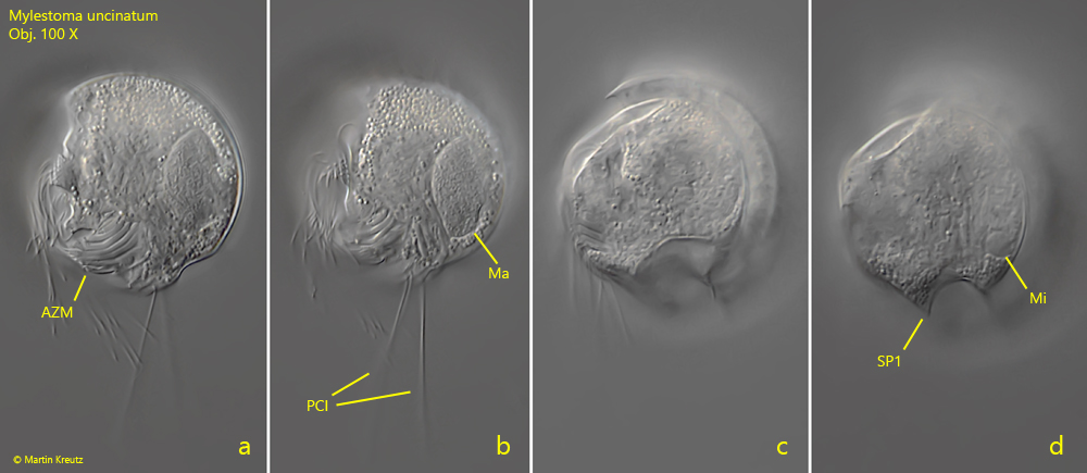
Fig. 6 a-d:Mylestoma uncinatum. L = 33 µm. Four focal planes of a specimen from the left side (a, b) to the right side (c, d). AZM = adoral zone of membranelles, MI. micronucleus, Ma = macronucleus, PCI = posterior cirri on left side, SP1 = posterior spine 1. Obj. 100 X.
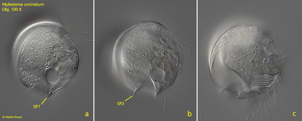
Fig. 7 a-c:Mylestoma uncinatum. L = 37µm. Three focal planes of the right side of a slightly squashed specimen. SP1 = tooth-shaped spine 1, SP2= spine pointing dorsally. Obj. 100 X.
Fig. 8 a-b:Mylestoma uncinatum. L = 31 µm. Two focal planes from ventral of a freely swimming specimen. Obj. 60 X.
Fig. 9 a-b:Mylestoma uncinatum. Two focal planes of a specimen during the process of cell division. The cells separate on the dorsal margins. The poor quality of the images is due to a high layer thickness. Obj. 100 X.