length 114–138 µm, 240–268 µm with terminal spines
head short and rounded
head with twao lateral, hyaline tentacles
two cephalic glands
three pairs of dorsal setolae on head
neck little constricted
posterior end with zwo fiinger-shapes appendages with short spines
a pair of 5–8 long spines ventrally
a pair of 3 long spines dorsally
dorsally and ventrally rhombic scales with short spines
Neogossea fasciculata
I find Neogossea fasciculata exclusively in the Simmelried. Most specimens are found in the siltation zone with very shallow water depths (< 10 cm) and large amounts of decomposing plant masses. So far, I have only found Neogossea fasciculata in the summer months from June to August, which is in contrast to the information provided by Schwank (1992), according to which Neogossea fasciculata prefers cooler temperatures.
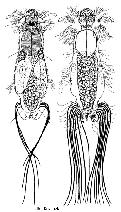
Neogossea fasciculata differs from the similar species Neogossea antennigera by the round head, the long terminal spines and above all by a pair of finger-shaped appendages at the posterior end, which bear a short, claw-shaped spine (s. figs. 3a and 16). The long spines at the posterior end were about 100 µm long in my specimens, whereas they are said to be only 10–35 µm long in Neogossea antennigera.
According to Krivanek (1959), of the long spines at the posterior end, a pair of each 3 spines should arise dorsally and a pair of each 5–8 spines on the ventral side. Remane (1936) found only one pair of each 6 spines on the ventral side (i.e. 12 spines in total). In my population the specimens had constantly a pair of each 8 spines on the ventral side (i.e. 16 in total, s. figs. 17 and 18). The number of terminal, elongated spines therefore appears to be variable.
The shape of the scales on both the ventral and dorsal sides differed considerably from Schwank’s description. According to this description, both the ventral and the dorsal scales should be rhombic in shape with short spines. According to my observations, the dorsal scales are shield-shaped, with a distal notch and a short, simple spine (s. fig. 14). Their length is 3–4 µm. In contrast, the ventral scales are very slender, have a comb-shaped keel and do not overlap (s. fig. 11). The keel has a short tip at the distal end. The scale also ends in a point, giving the impression of a distal double point (s. fig. 12).
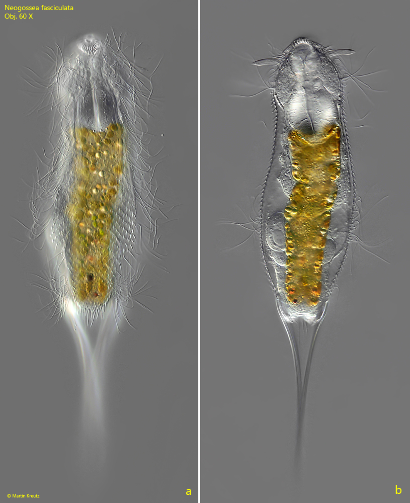
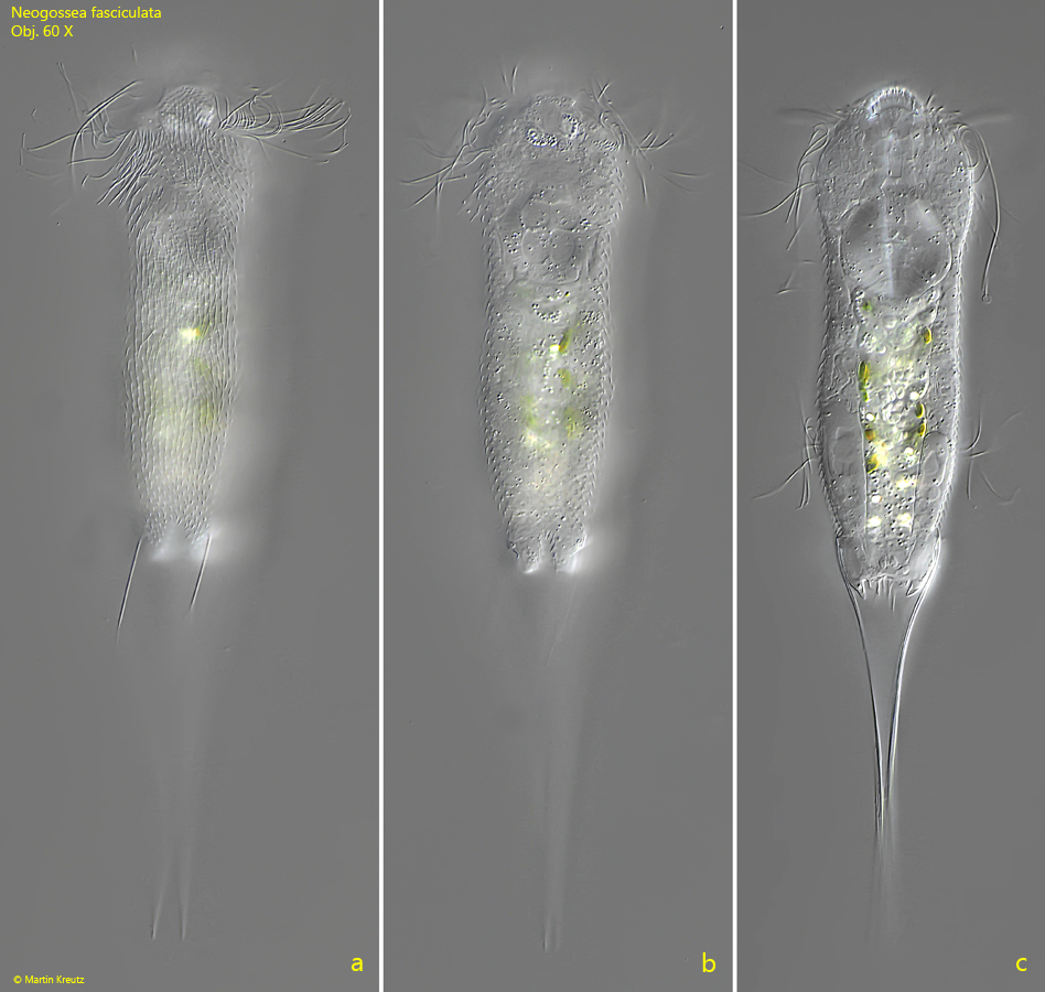
Fig. 1 a-b:Neogossea fasciculata. L = 234 µm (with terminal spines). A freely swimming specimen from ventral. Obj. 60 X.
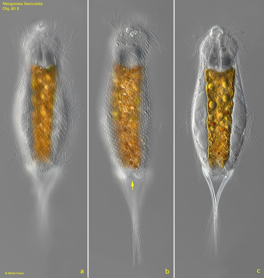
Fig. 2 a-c:Neogossea fasciculata. L = 245 µm (with terminal spines). A second, freely swimming specimen from dorsal. Note the terminal incision of the body (arrow). Obj. 60 X.
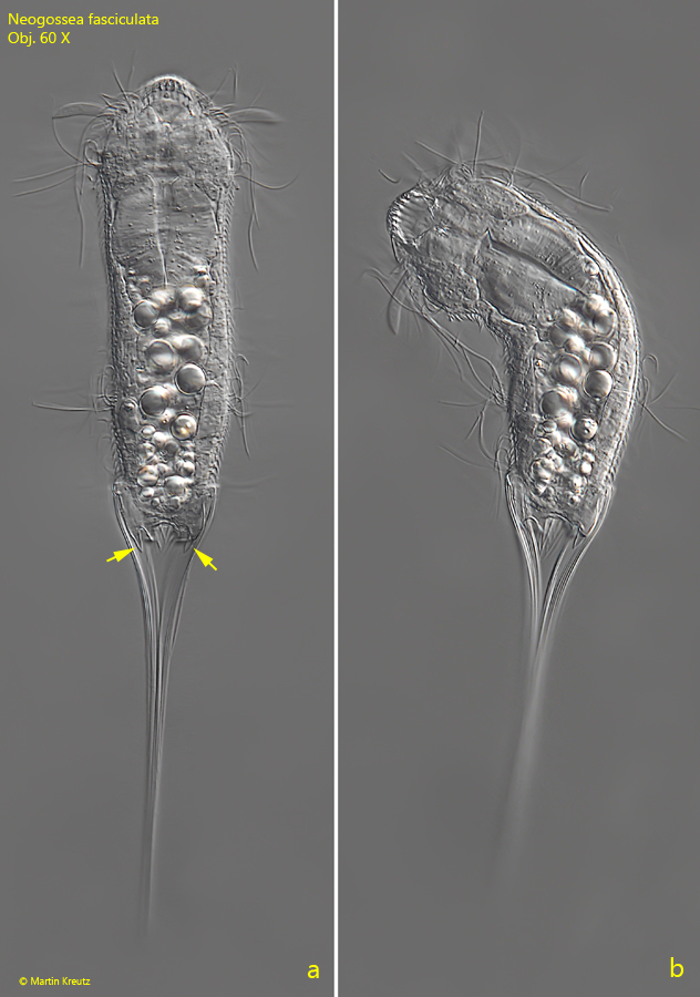
Fig. 3 a-b:Neogossea fasciculata. L = 226 µm (with terminal spines). A third, likely young specimen from ventral. Note the finger-shaped appendages with a short spine at the distal end (arrows). Obj. 60 X.
Fig. 4 a-c:Neogossea fasciculata. L = 210 µm (with terminal spines). A fourth specimen from dorsal. Obj. 60 X.
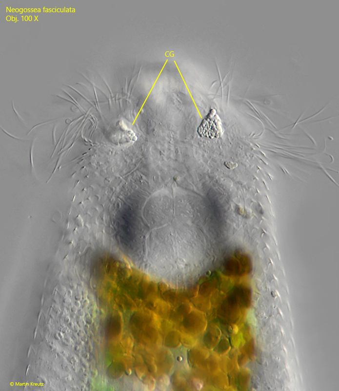
Fig. 5:Neogossea fasciculata. The pair of cephacil glands (CG) at the dorsal side of the head. The function of the glands is unknown. Obj. 100 X.
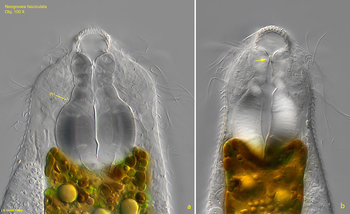
Fig. 6 a-b:Neogossea fasciculata. The pharynx (PH) in a squashed (a) and slightly squashed specimen (b). Note the two flaps (arrow) that close the pharynx to the mouth opening. Obj. 100 X.
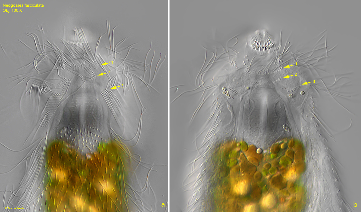
Fig. 7 a-b:Neogossea fasciculata. The pattern of the ciliation (a) and of the basal bodies (b) in 3 rows of the ventral side of the head (a). Obj. 100 X.
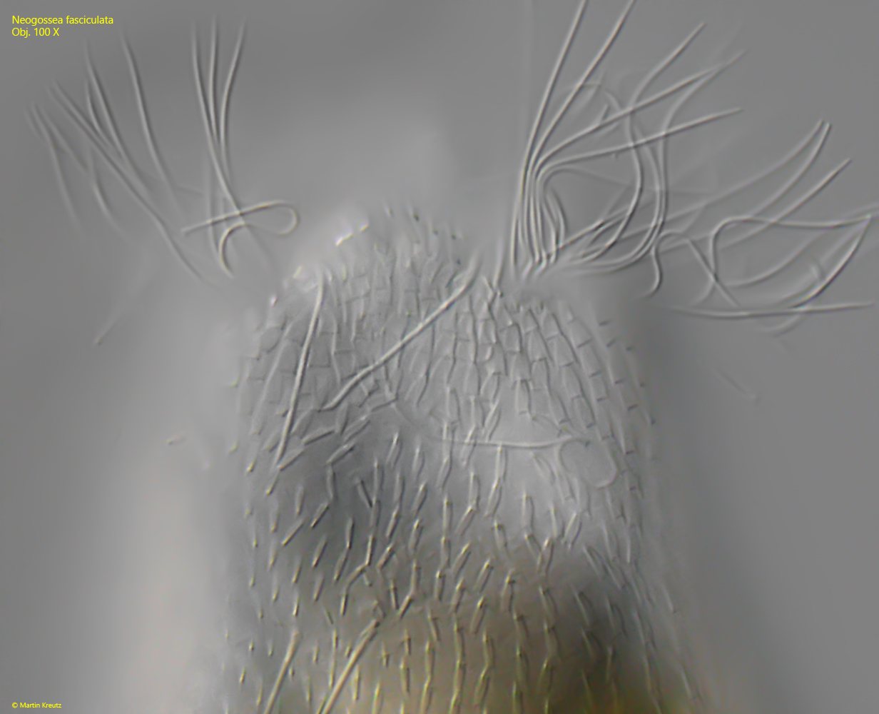
Fig. 8:Neogossea fasciculata. The scales of the dorsal side of the head. Obj. 100 X.
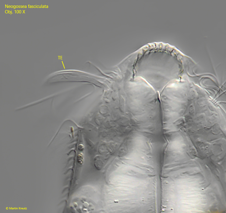
Fig. 9:Neogossea fasciculata. The right tentacle of a specimen in detail. The tentacle has a length of 18 µm and is a protrusion of the cuticle. A fine filament runs through it, which could be a nerve fiber. This would indicate a sensory function. Obj. 100 X
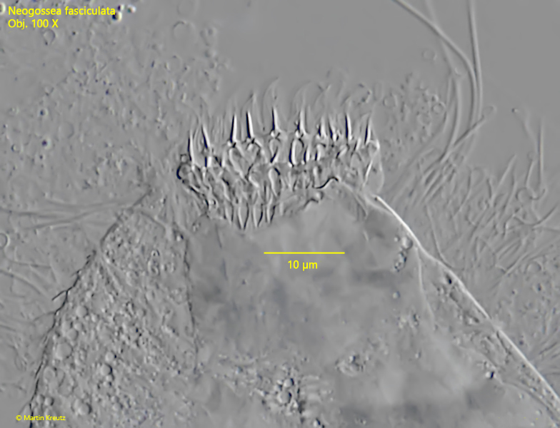
Fig. 10:Neogossea fasciculata. The distal end of the mouth tube is a ring of fine spines. Obj. 100 X
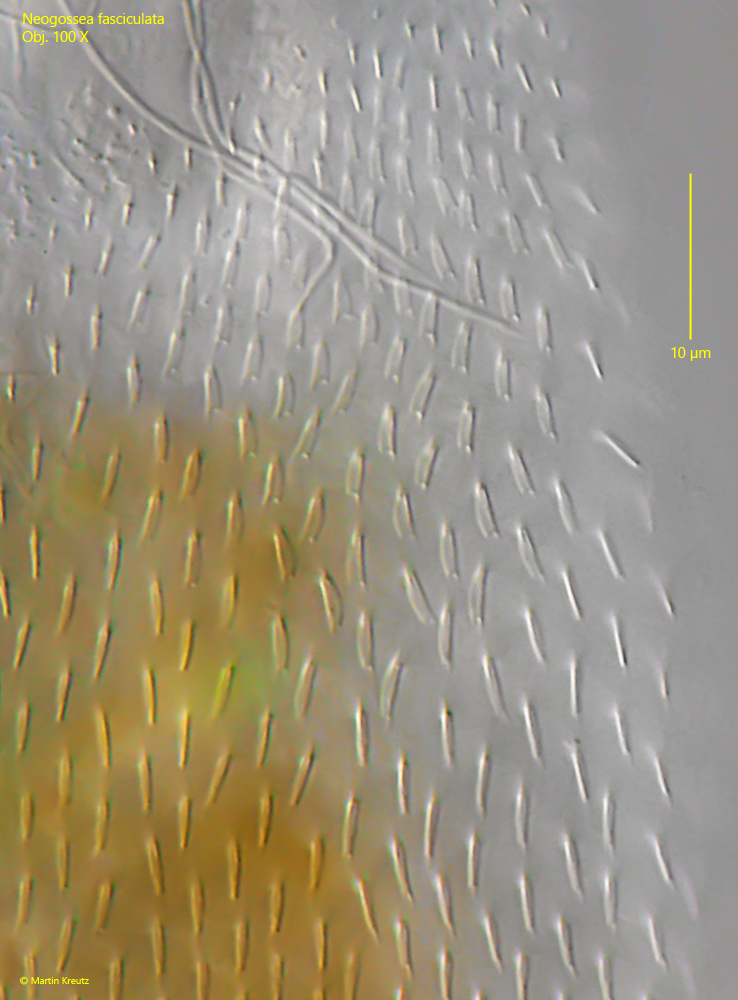
Fig. 11:Neogossea fasciculata. The slim ventral scales are 3–4 µm long and keeled. Obj. 100 X
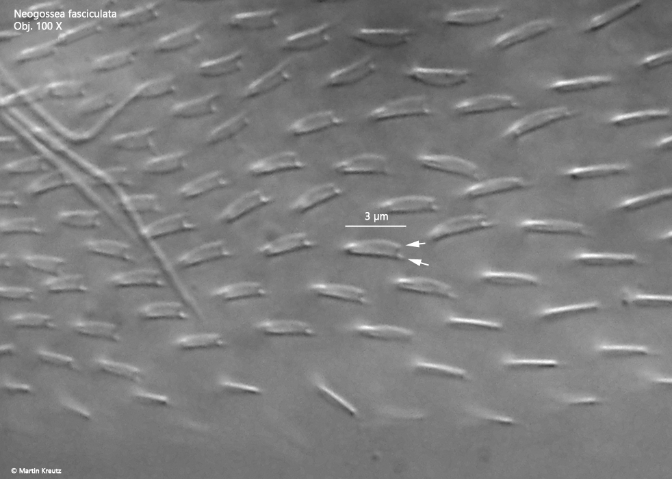
Fig. 12:Neogossea fasciculata. The ventral scales in detail. The keel and the scale each end in a point at the distal end, creating a double point (arrows). Obj. 100 X.
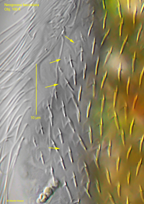
Fig. 13:Neogossea fasciculata. Lateral viev of the dorsal scales with a short, straight spine. Obj. 100 X.
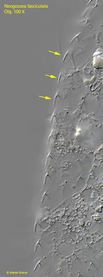
Fig. 14:Neogossea fasciculata. The dorsal scales are 3–4 µm long, shield-shaped (arrows) with a distal incision and a short, simple spine. Obj. 100 X
Fig. 15:Neogossea fasciculata. The elongated scales at the posterior end of the ventral side. Obj. 100 X
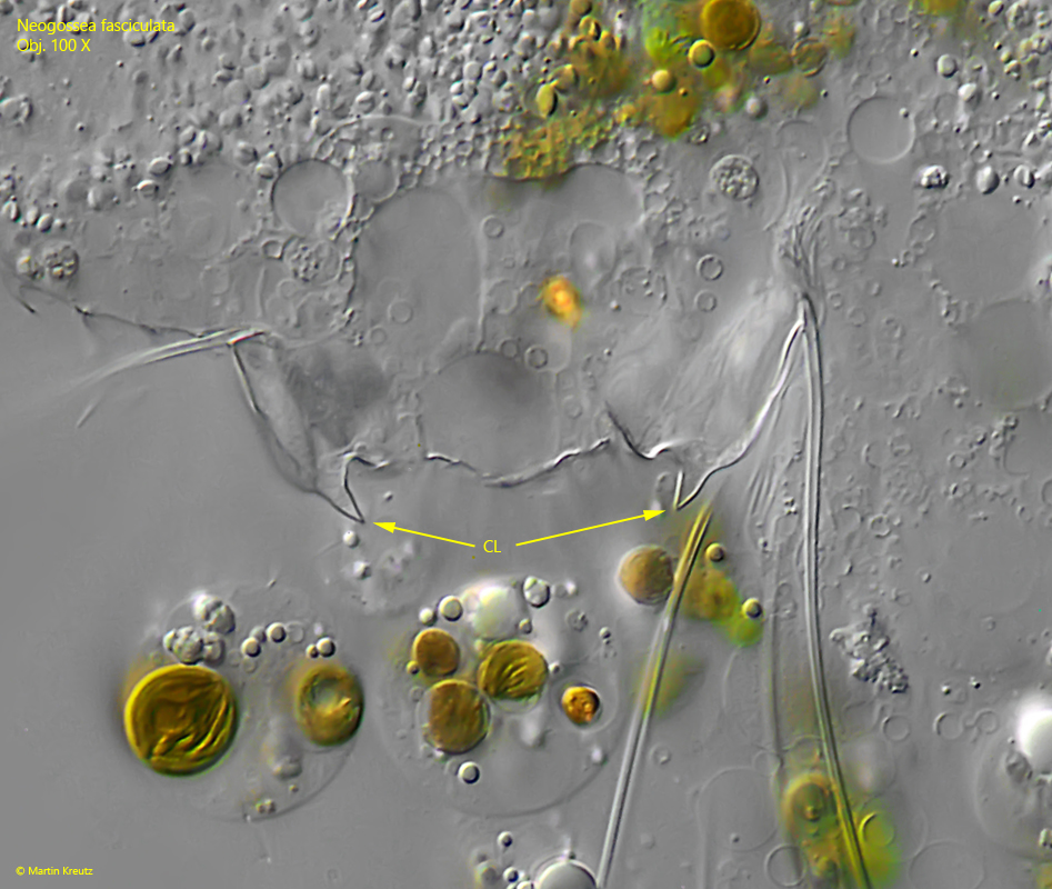
Fig. 16:Neogossea fasciculata. The the posterior appendages with claw-shaped spines (CL) at the distal end. Obj. 100 X
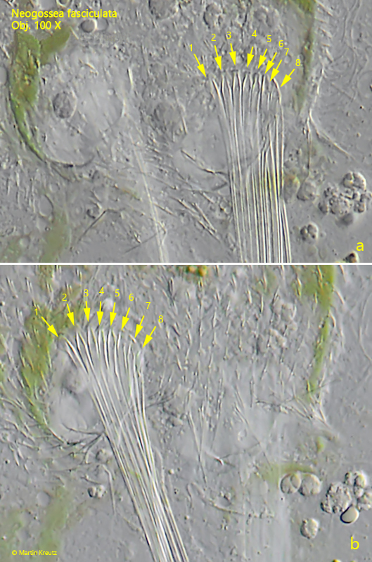
Fig. 17:Neogossea fasciculata. The bases of the terminal spines from ventral on the left side (a) and right side (b). On each side 8 spines arise. Obj. 100 X
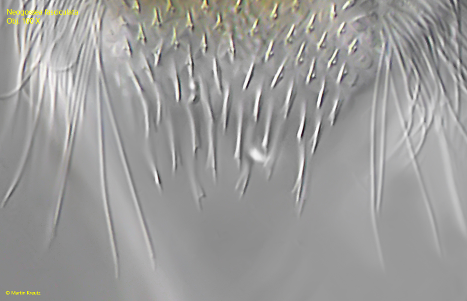
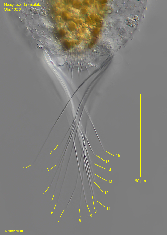
Fig. 18:Neogossea fasciculata. The 16 elongated spines at the posterior end with a maximum length of 100 µm (spine 7). Obj. 100 X
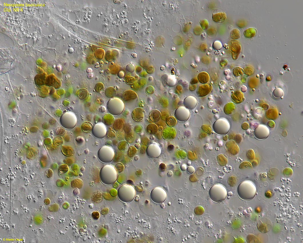
Fig. 19:Neogossea fasciculata. The content of the intestine in a strongly squashed specimen. The diet seems to consist of Chrysophyceae, Cryptomonads and Rhodobacteria. Obj. 100 X