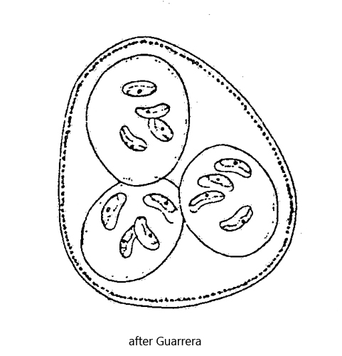
daughter cells remain united within gelatinized cell wall of mother cell
cells 10–20 µm long, kidney-shaped, slightly curved with rounded apices
one pyrenoid
one parietal chloroplast
planctonic lifestyle
Nephrocytium limneticum
I find Nephrocytium limneticum very rarely in plankton samples, always with several years between them. Nephrocytium can be confused with Kirchneriella, whose cells are also kidney-shaped or crescent-shaped. However, the colonies of Nephrocytium are covered by a distinct and thick layer of mucus, which is absent in Kirchneriella. In addition, the daughter colonies of Nephrocytium remain united for a long time in the swollen and gelatinized cell wall of the mother cell (s. fig. 2 a).
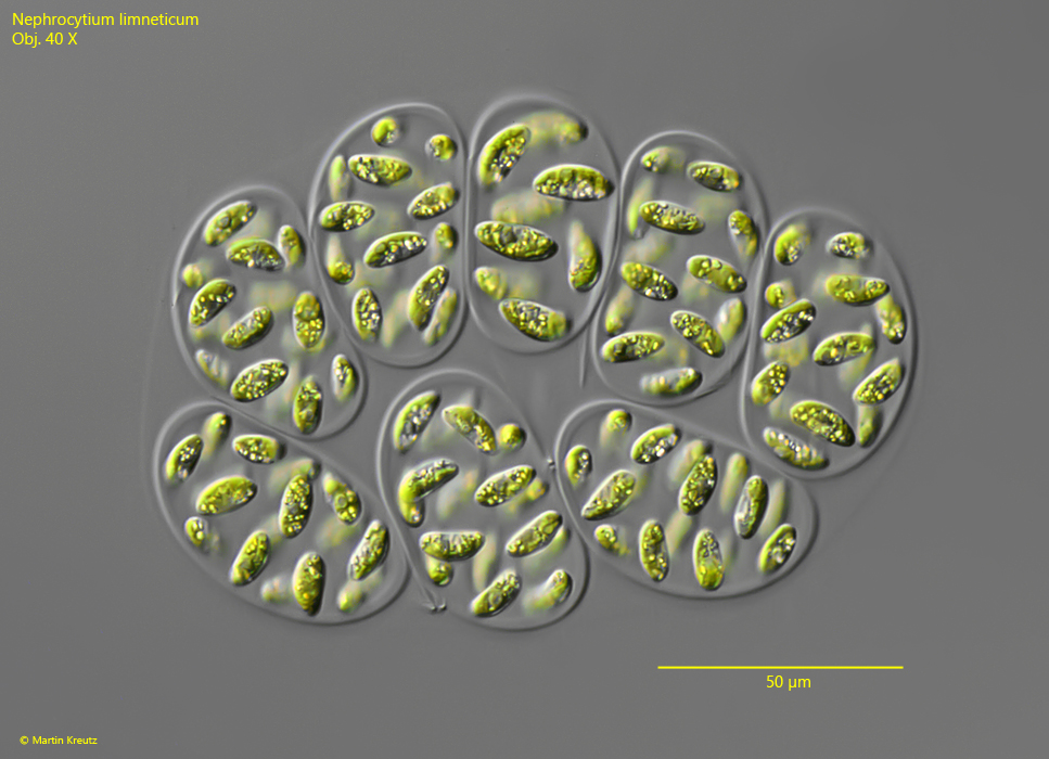
Fig. 1:Nephrocytium limneticum. L = 156 (of coenobium). A coenobium of 8 colonies with 16 cells each within the gelatinized cell wall of the mother cell. Obj. 40 X.
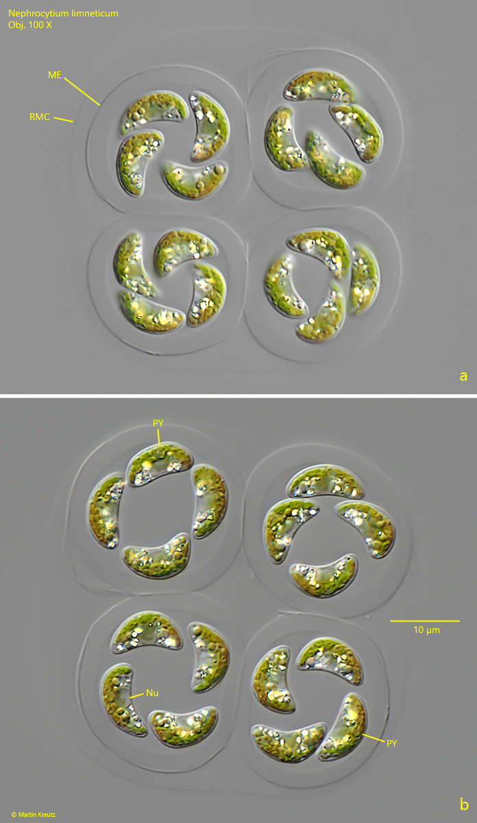
Fig. 2 a-b:Nephrocytium limneticum. L = 10–12 µm (of the cells). A slightly squashed (a) and a strongly squashed (b) coenobium of 4 colonies with 4 cells each within the remains of the mother cell (RMC). Each colony is in a thick mucilage envelope (ME). Nu = nucleus, PY = pyrenoid. Obj. 100 X.
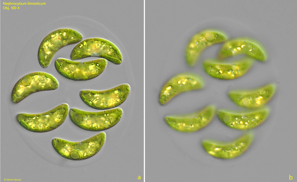
Fig. 3 a-b:Nephrocytium limneticum. L = 19–21 µm (of the cells). Two focal planes of a second coenobium of 8 cells. Obj. 100 X.