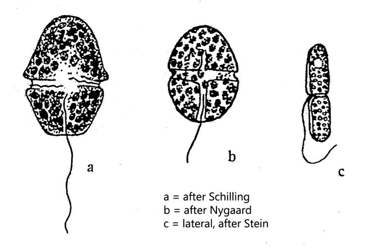
epicone (anterior half) longer than hypocone (posterior half)
eyespot sometimes present
chloroplasts numerous, blue-green
Nusuttodinium aeruginosum
I could detect Nusuttodinium aeruginosum (previously Gymnodinium aeruginosum) up to now exclusively in Ulmisried. The species can be identified easily by the blue-green color. In fact, Nusuttodinium aeroginosum is a colorless species. The chloroplasts, which give the cell its blue-green coloration, were “stolen” from photosynthetic prey organisms. Such stolen chloroplasts are also called cleptoplasts. It could be proved that Nussuttodinium aeroginosum obtains these kleptoplasts from Chroomonas. The cells of the prey organisms are completely digested except for the chloroplasts. These are then incorporated into the peripheral cell layer and kept alive.
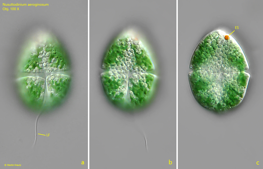
In the literature, the species is described without an eyespot. I could detect an eyespot in some specimens (s. Fig. 1 and Fig. 5). Thus, the species seems to be quite variable not only with respect to the cell shape.
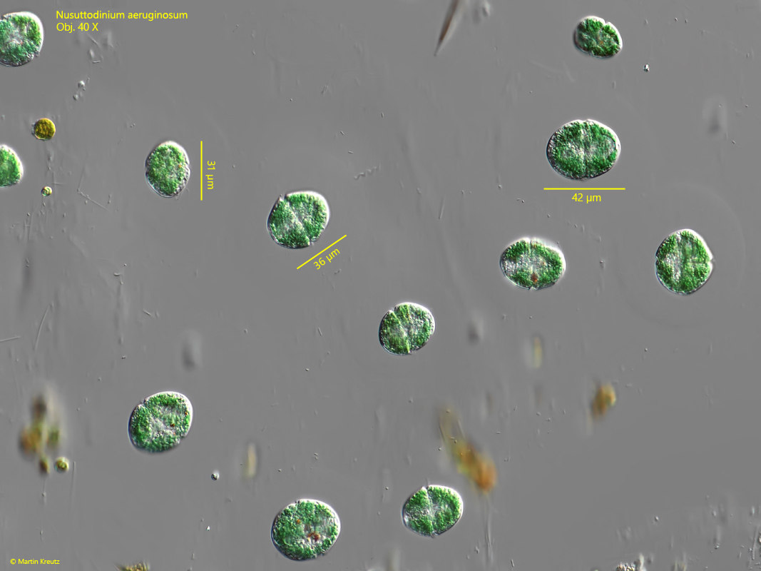
Fig. 1: Nusuttodinium aeruginosum. Overview of a mass occurrence. Note the two cells with an eye spot. Obj. 40 X.
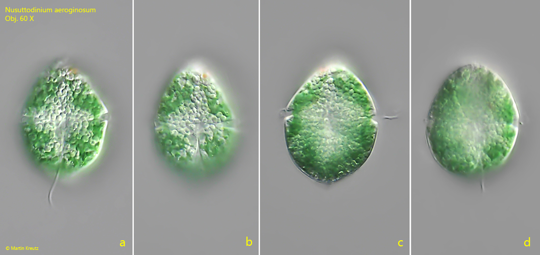
Fig. 2 a-d: Nusuttodinium aeruginosum. L = 37 µm. Different focal planes of a freely swimming specimen from ventral. Obj. 60 X.
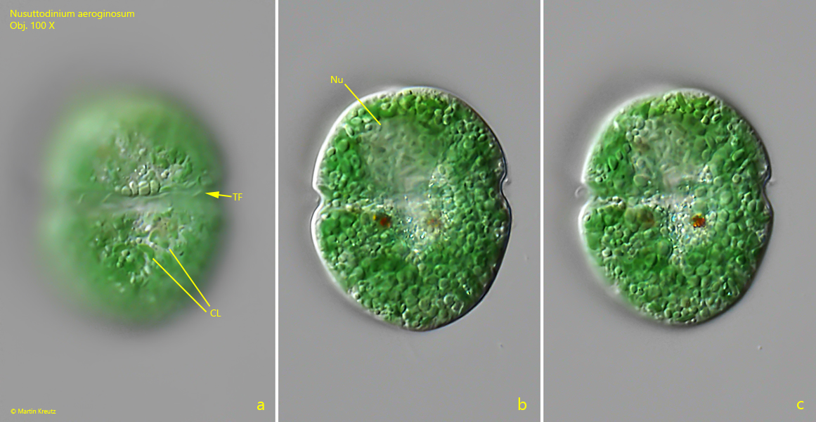
Fig. 3 a-c: Nusuttodinium aeruginosum. L = 38 µm. A slightly squashed specimen from dorsal. CL = cleptoplasts, NU = nucleus, TF = transversal flagellum. Obj. 100 X.
Fig. 4 a-c: Nusuttodinium aeruginosum. L = 36 µm. A slightly squashed specimen from ventral. ES = eyespot, LF = longitudinal flagellum. Obj. 100 X.