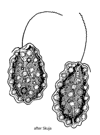
single chloroplast, brown-yellowish, without pyrenoid
cell surface with conspicuous blisters (mucucysts)
nucleus in anterior third
Ochromonas verrucosa
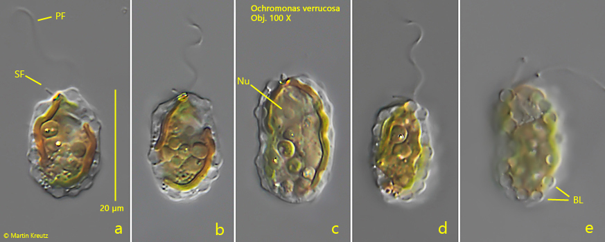
I find Ochromonas verrucosa rarely but regularly in some of my sites. Mostly in samples from the water surface between floating plants or decomposing plant masses. In a few cases I could also observe mass development in spring. The species can be easily identified by the golden brown color caused by fucoxanthin and the typical blisters (mucocysts) on the cell surface. Mostly the cells are oval shaped. Since they are slightly metabolic, they can also stretch in length. Ochromonas verrucosa is often associated with the similar species Ochromonas crenata. However, Ochromonas crenata is smaller and has an almost spherical body shape.
Fig. 1 a-e: Ochromonas verrucosa. L = 20 µm. A freely swimming specimen. Note the characteristic blisters (BL, fig. 1e) on the cell surface. Nu = nucleus, PF = primary flagellum, SF = secondary flagellum. Obj. 100 X.