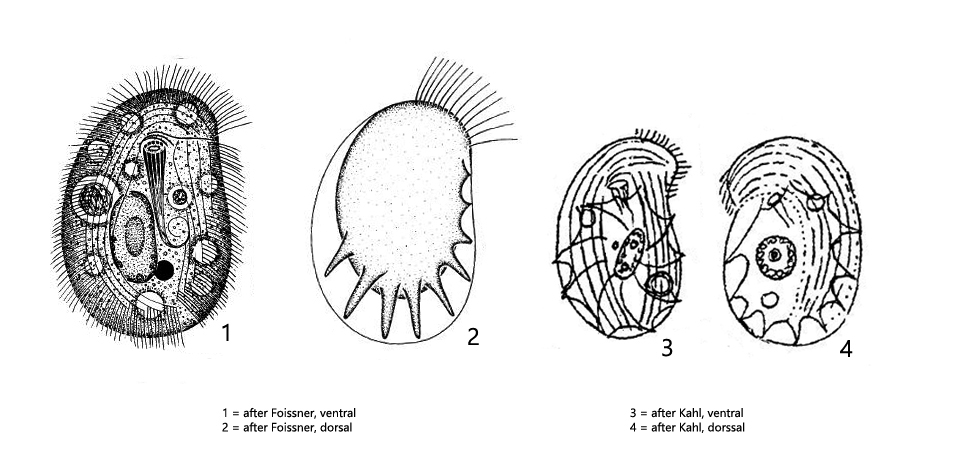
body shape oval with an anterior left-hand rostrum
length 30–50 µm, width 25–35 µm
dorso-ventrally flattened
posterior margin of the dorsal side with 2–10 radiating spines
cytophyarynx with pharyngeal basket on ventral side in anterior third
somatic kineties of the ventral side from Chilodonella type with a field devoid of cilia in the middle
dorsal brush parallel to anterior, left margin
two diagonally arranged contractile vacuoles
one ellipsoidal or oval macronucleus with one adjacent micronucleus
Odontochlamys gouraudi
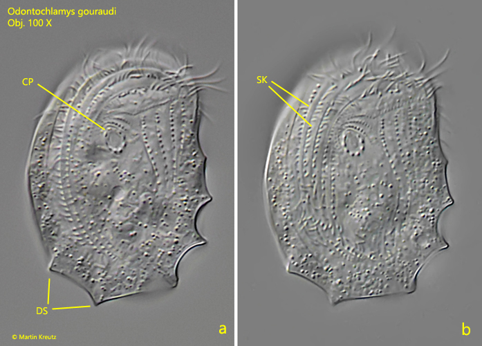
Odontochlamys gouraudi is a ciliate of highly peculiar shape that I find in moss samples. To extract Odontochlamy gouraudi from the moss, I crush some moss (e.g. taken from a tree trunk or stone) and soak the small pieces in some rainwater in a petri dish. On the water surface I put some floating coverslips. After only a few days Odontochlamys gouraudi colonizes the coverslips, along which it glides with its ventral side. It is thus very easy to observe. The radiating spines of the dorsal side, which protrude the posterior margin, can be easily seen even at low magnifications. Because of this feature Odontochlamys gouraudi cannot be confused with any other species. Apart from this character, the ventral side looks very similar to the genus Chilodonella (s. figs. 1 a-b and 2 a-b). In the middle of the ventral side there is a cilia-free field. The dorsal side is naked except for a single-row dorsal brush, which follows the anterior left margin and is also visible from ventral (s. fig. 3).
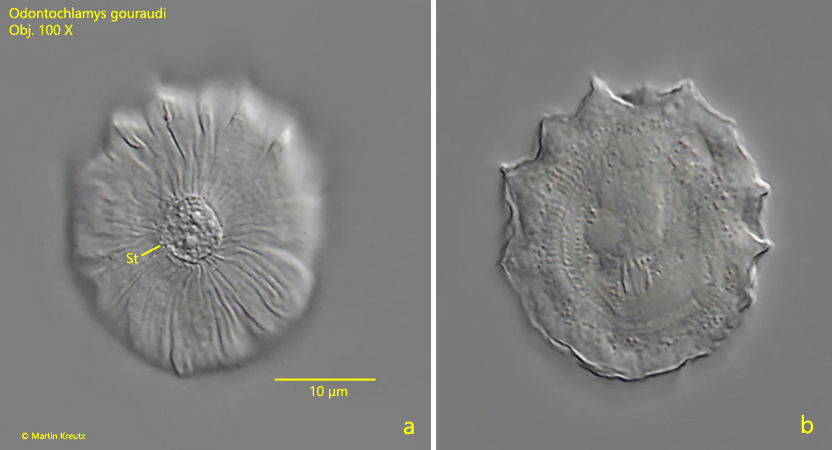
In older samples I could find numerous cysts of Odontochlamys gouraudi on the floating coverslips (s. fig. 4). They are easy to identify because the typical dorsal spines are still visible in the cysts. The cysts had a diameter of about 25 µm. They were attached to the coverslip with a very short stalk.
Fig. 1 a-b:Odontochlamys gouraudi. L = 37 µm. Two focal planes of the ventral side of a freely gliding specimen. CP = cytopharynx, DS = dorsal spines, SK = somatic kineties of the ventral side. Obj. 100 X.
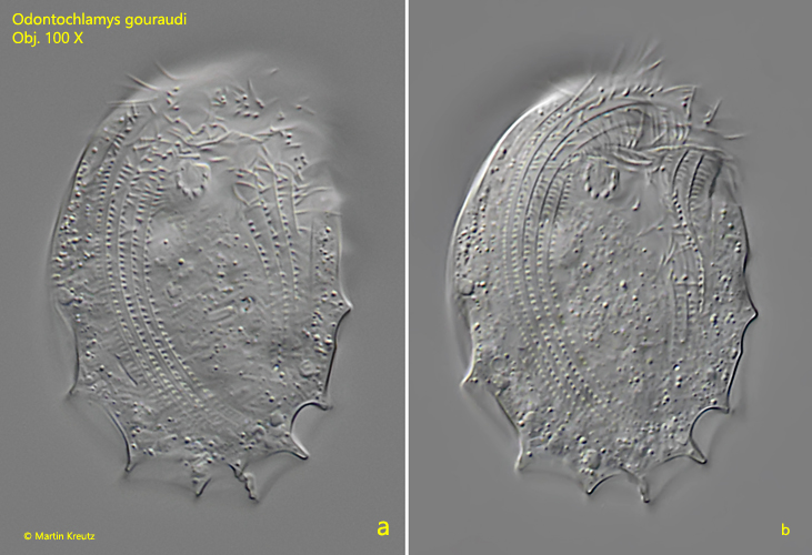
Fig. 2 a-b:Odontochlamys gouraudi. L = 44 µm. Two focal planes of the ventral side of a second specimen. Obj. 100 X.
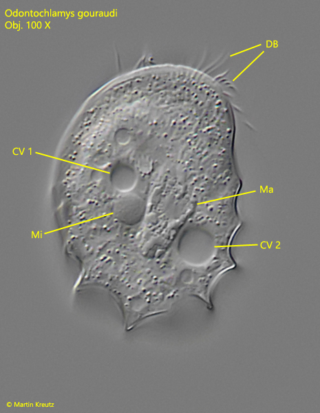
Fig. 3:Odontochlamys gouraudi. L = 37 µm. Focal plane on the two, diagonally arranged contractile vacuoles (CV 1 , CV 2) the macronucleus (Ma) and the adjacent micronucleus (Mi). DB = dosal brush. Obj. 100 X.
Fig. 4:Odontochlamys gouraudi. Two focal planes of a cyst with a diameter of 25 µm. St = stalk. Obj. 100 X.