body ovoid or elongated oval, slightly asymmetric, metabol
length 25–50 µm (commonly 30–40 µm)
12–14 longitudinal rows of cilia
spindle-shaped extrusomes, about 0.5 µm long
cytopharynx apical, slightly sub-polar
globular macronucleus central
one spherical micronucleus adjacent to macronucleus
contractile vacuole terminal and sub-polar
one caudal cilium
Paraurotricha discolor
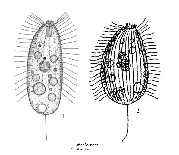
Paraurotricha discolor was first described by Kahl (1930) as Urotricha discolor. In 1983 Foissner published a redescription of the species and transferred it to the newly created genus Paraurotricha on the basis of morphological studies.
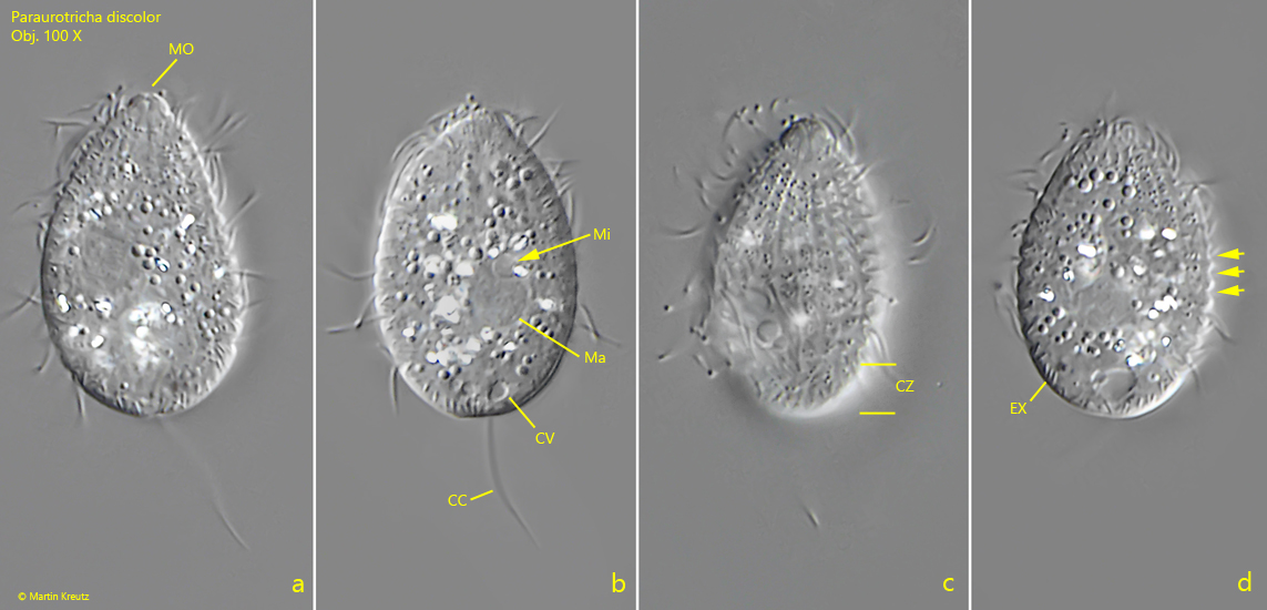
I have so far only found Paraurotricha discolor in the Simmelried. However, as the species is common, I may have overlooked it in my other sampling sites. In contrast to representatives of the genus Urotricha, the body of Paraurotricha discolor is very flexible and the ciliate burrows in detritus. The size is given by Foissner as 30–40 µm and by Kahl as 40–50 µm. However, Kahl mentions he having found specimens with a length of 25 µm. The size seems to be quite variable. The cilia stand in small depressions, which is why the edge of the pellicle appears notched (s. fig. 1 d). The posterior quarter of the body is naked, as in Urotricha (s. fig. 1 c). According to Foissner, the extrusomes should be straight and spindle-shaped with a length of about 0.5 µm. According to my measurements they are about 1 µm long. In terms of body shape, my specimens were more similar to Kahl’s drawing (s. drawing 2, above). I have not yet found such slender specimens as Foissner drew them (s. drawing 1 , above). However, I found the contractile vacuole to be terminal, as Foissner it indicates, and not sub-terminal, as Kahl drew them (s. drawing 2, above).
Fig. 1 a-d: Paraurotricha discolor. L = 26 µm. Different focal planes of a freely swimming specimen. Note the cilia free zone (CZ) at the posterior end and the notched pellicle (d, arrows). CC = caudal cilium, CV = contractile vacuole, EX = extrusomes, Ma = macronucleus, Mi = micronucleus, Mo = mouth opening. Obj. 100 X.