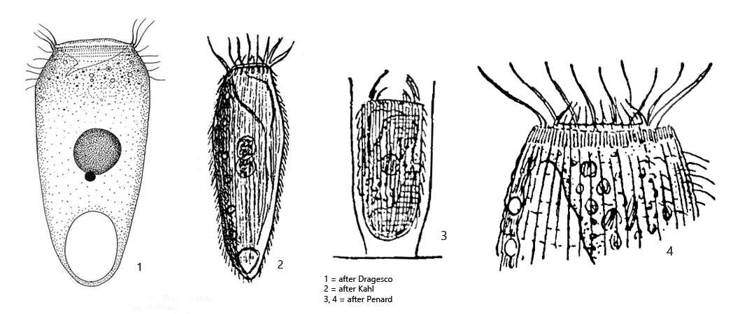
posterior end tapered, broadly rounded or slightly pointed
length 120–220 µm
circular oral aperture with large, permanent cytopharynx (receptaculum)
a large, non-contractile vacuole at posterior end, connected with permament cytopharynx
numerous small contractile vacuoles distributed over midbody
macronucleus globular in midbody with a large adjacent micronucleus
lorica membranous, cylindrically shaped
Pelatractus grandis
I have found Pelatractus grandis so far exclusively in the Simmelried. It lives in the upper mud zone and is associated for example with metopid ciliates. There are only few descriptions of Pelatractus grandis available. Kahl copied most of the original description from Penard. He found only one specimen which he could examine. Later an examination on fixed material was done by Dragesco et al. (1974) and a silver impregnation. Further descriptions are not available to my knowledge.
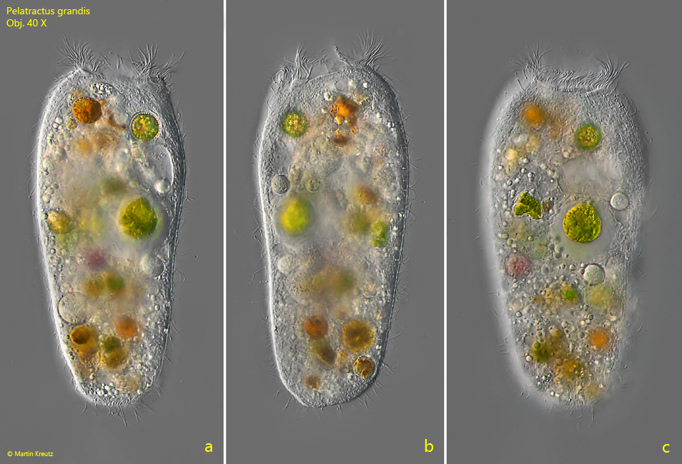
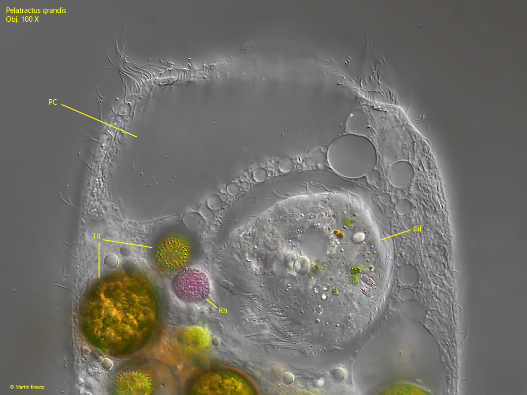
Pelatractus grandis is quite stately and grows about 180–200 µm long in my population. The freely swimming specimens were mostly broadly rounded at the posterior end and never as pointed as Kahl has drew them (s. drawing 2 above and fig. 1 a-c). The anterior end is truncated transversely and is formed by the circular peristome. Adjacent to this is a large cavity called the permanent cytopharynx. Kahl has chosen the term receptaculum for it. This permanent cytopharynx is thought to be associated with the terminal vacuole. This terminal vacuole is not the contractile vacuole. I could clearly recognize it in my population (s. fig. 3). However, I could not recognize the connection to the permanent cytopharynx. The actual contractile vacuoles are numerous and distributed mainly over the midbody region (s. figs. 5 and 6).
Pelatractus grandis seems to feed on anything that fits through the cytopharynx. In the food vacuoles I could detect a wide variety of prey organisms such as Trachelomonas, rhodobacteria, ciliates, flagellates, algae and dinoflagellates (s. fig. 9).
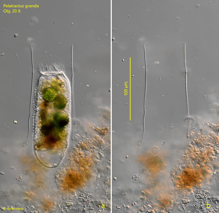
The macronucleus is globular and has a closely adjacent, relatively large micronuleus (s. fig. 7). Strangely, Kahl seems to have drawn two macronuclei (s. drawing 2, above) and does not mention the nuclear apparatus in his description either. However, Kahl’s description mentions the lorica that Pelatractus grandis builds. It is described to be beaker-shaped. So far I could find only one specimen in its lorica (s. fig. 4 a-b). It is cylindrical with smooth, parallel walls and about as long as the ciliate that built it. The lorica is left upon even slight disturbances. Therefore the images in fig. 4 a-b were taken without coverslip.
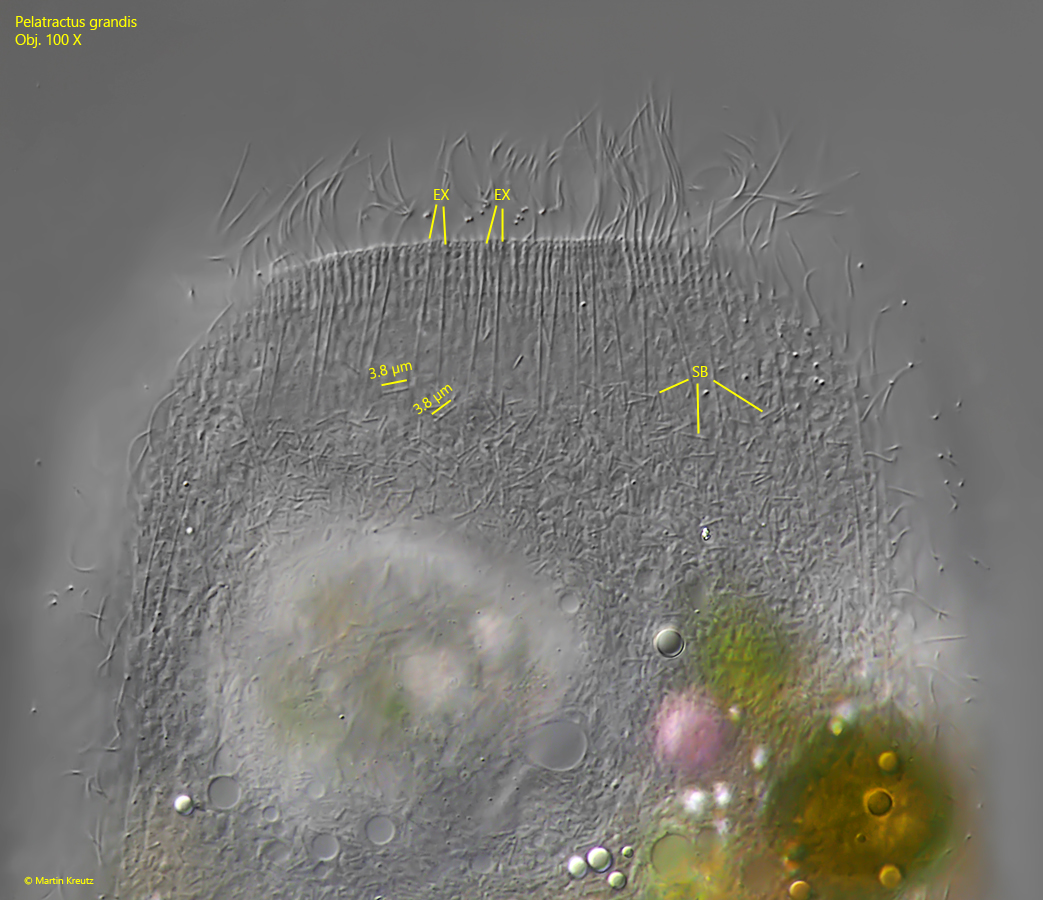
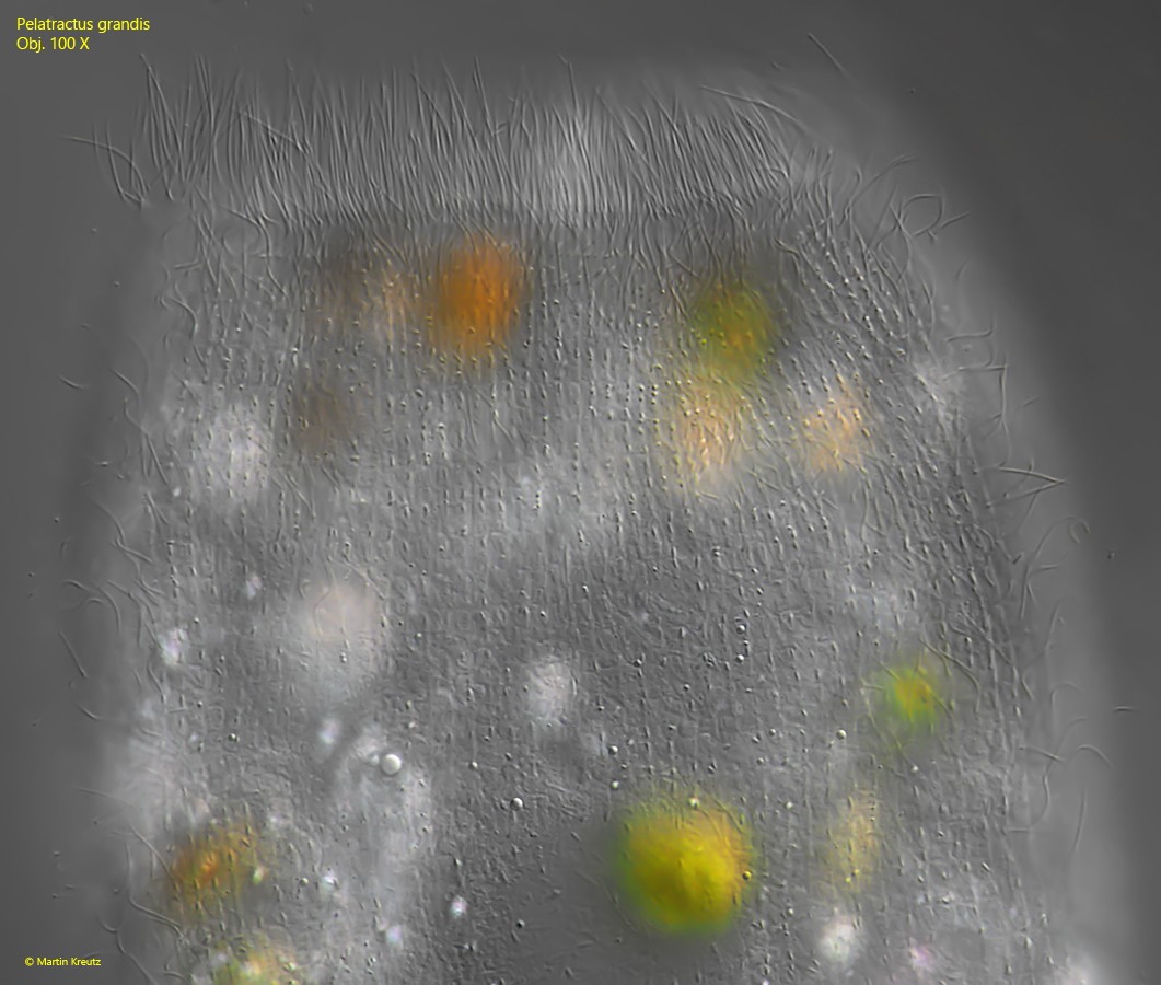
The edge of the persistom is provided with thin, rod-shaped extrusomes. According to my observations they are arranged in pairs (s. fig. 8). Possibly, however, they are also trichites, which should mechanically stabilize the cytopharynx. In the cytoplasm I could detect large amounts of symbiotic bacteria (s. fig. 8), which are not mentioned by previous authors. They are distributed throughout the cytoplasm. They seems to be only one kind of rod-shaped bacteria, which are constantly 3.8 µm long. The surface of the pellicle has a very light square pattern, as known from the related species Vasicola ciliata (s. fig. 10).
Fig. 1 a-c:Pelatractus grandis. L = 187 µm. A freely swimming specimen. Obj. 40 X.
Fig. 2 a-c:Pelatractus grandis. L = 190 µm. A second freely swimming specimen. Obj. 60 X.
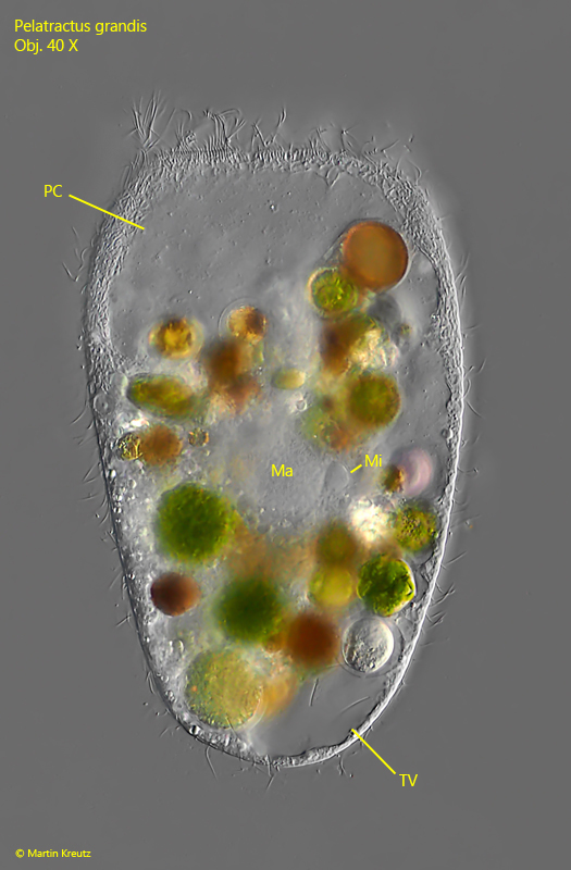
Fig. 3:Pelatractus grandis. L = 184 µm. A slightly squashed specimen. Note the large permanent cytopharynx (PC, called receptaculum by Kahl) and the terminal vacuole (TV) connected to the permanent pharynx. The terminal vacuole is not the contractile vacuole. Ma = macronucleus, Mi = micronucleus. Obj. 40 X.
Fig. 4 a-b:Pelatractus grandis. L = 178 µm. A specimen in the membranous, cylindrically shaped lorica (a) and the empty lorica (b) after the specimen has left. The lorica has a length of 185 µm und the aperture is 75 µm wide. Obj. 20 X.
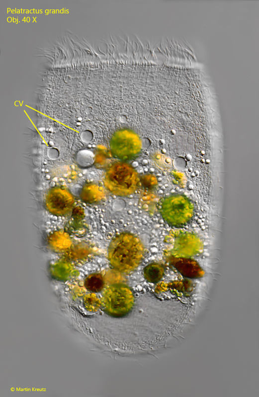
Fig. 5:Pelatractus grandis. L = 193 µm. Focal plane on the numerous contractile vacuoles (CV) distributed over the midbody. Obj. 40 X.
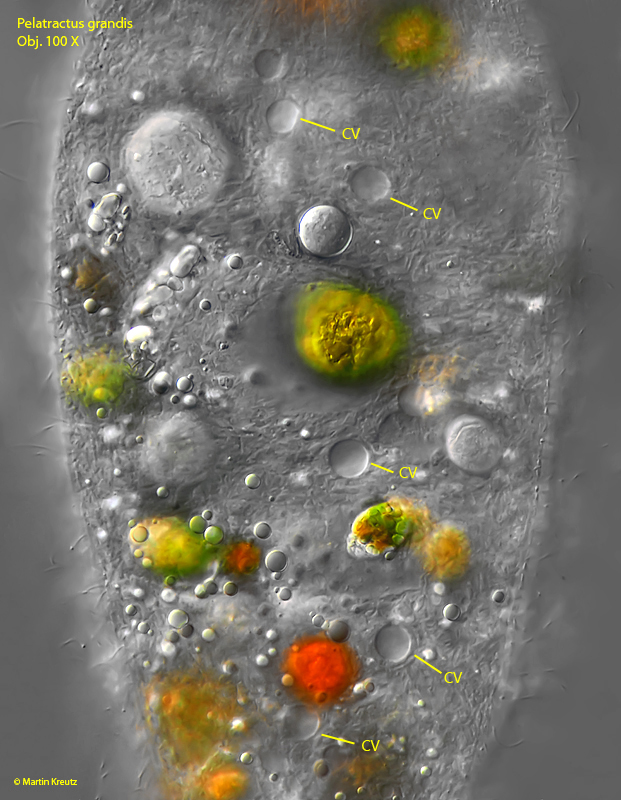
Fig. 6:Pelatractus grandis. A closer view of the contractile vacuoles (CV) in a slightly squashed specimen. Obj. 100 X.
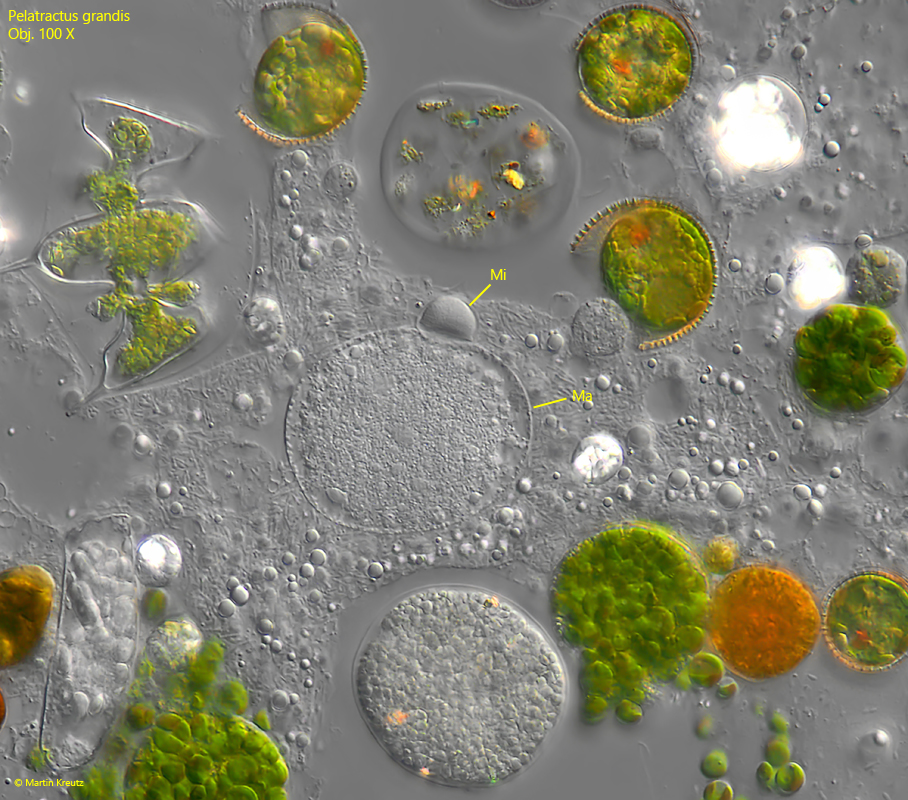
Fig. 7:Pelatractus grandis. The globular macronucleus (Ma) with the adjacent micronucleus (Mi). Obj. 100 X.
Fig. 8:Pelatractus grandis. Focal plane on the peristome. The edge of the cytopharynx appears to be equipped with thin, rod-shaped extrusomes (EX) arranged in pairs. In addition, large amounts of symbiotic bacteria (SB) are visible in the cytoplasm. These are thin rods with a constant length of 3.8 µm. Obj. 100 X.
Fig. 9:Pelatractus grandis. The same specimen as shown in fig. 6 with focal plane on the food vacuoles. This specimen preyed on Trachelomonas (TR), Rhodobacteria (Rh), and a large ciliate (Cil). PC = permanent cytopharynx. Obj. 100 X.
Fig. 10:Pelatractus grandis. Focal plane on the surface of the pellicle. A very faint pattern of squares can be recognized. Obj. 100 X.