cells spindle-shaped, cylindrical or sac-shaped, metabolic
length 22–81 µm
periplast striated with small rods parallel to striation
locomotion flagellum 1–1.5 X of body length
only distal end of locomotion flagellum moving during swimming
trailing flagellum with body length, attached to the ventral side in a groove
flagellar canal with a slit-like opening that extends from the apex backwards for about
trailing flagellum arises from a lateral slit of the mouth opening
ingestion apparatus with two rods
nucleus spherical
contractile vacuole at anterior end near ingestion apparatus
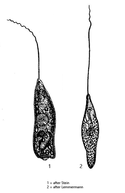
Peranema trichophorum
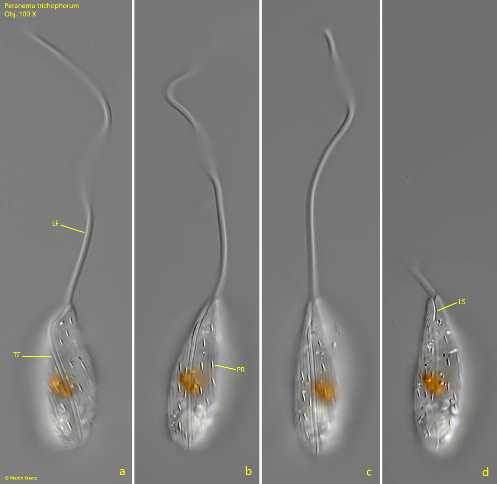
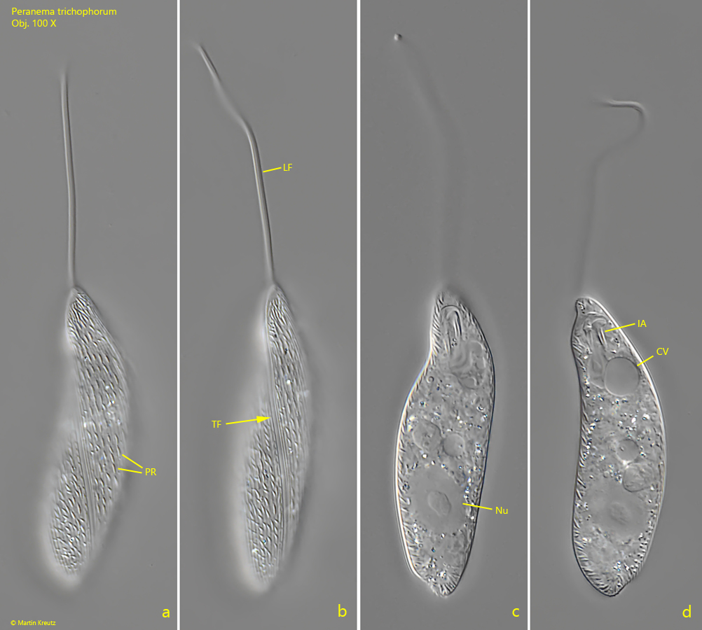
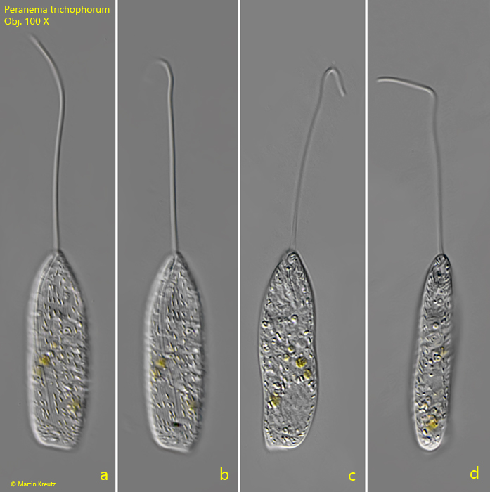
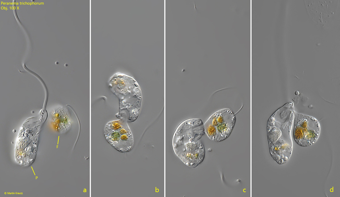
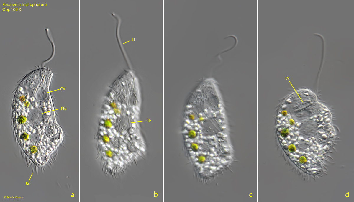
I find Peranema trichophorum in all my sites. This flagellate, which belongs to the Euglenophyceae, can be observed especially well on the floating cover glass, on which it often settles. It glides along the cover glass with a straight locomotion flagellum. The locomotion flagellum is described to be 1–1.5 X of the the body length (s. fig. 2 a-d). However, in my population I found specimens that had a locomotion flagellum twice of the body length (s. fig. 1 a-d). When swimming, Peranema trichophorum is often sac-shaped and one corner of the posterior end often protruding (s. fig. 3 a-d). The pellicle is distinctly striated and parallel to these stripes small rods are arranged of unknown function. In May 2014 I found a specimen in which these rods were assembled on the outside of the pellicle, giving the impression that the specimen was covered with bristles (s. fig. 5 a-d). In the literature I could not find any hint that these bristlest has been described before. Peranema trichophorum feeds heterotrophically and phagocytizes other protozoans. On one occasion I could observe Peranema trichophorum attacking a flagellate and tried to ingested it (s. fig. 4 a-d). The rods of the ingestion apparatus serves to widen the mouth opening and to bend back the locomotion flagellum. The attacked flagellate was obviously too large and was finally not phagocytosed.
Fig. 1 a-d:Peranema trichophorum. L = 28 µm (without flagellum). A freely swimming specimen. Note the lateral slit (LS) of the mouth opening where the trailing flagellum (TF) arise. LF = locomotion flagellum, PR = pellicular rods. Obj. 100 X.
Fig. 2 a-d:Peranema trichophorum. L = 55 µm (without flagellum). A second, larger specimen. CV = contractile vacuole, IA = ingestion apparatus, LF = locomotion flagellum, Nu = nucleus, PR = pellicular rods, TF = trailing flagellum. Obj. 100 X.
Fig. 3 a-d:Peranema trichophorum. L = 50 µm (without flagellum). A third, freely swimming specimen. Obj. 100 X.
Fig. 4 a-d:Peranema trichophorum. A flagellate (F) is attacked by Peranema trichophorum (P). Obj. 100 X.
Fig. 5 a-d:Peranema trichophorum. L = 52 µm (without flagellum). A slightly squashed specimen covered with bristles (Br). CV = contractile vacuole, IA = ingestion apparatus, LF = locomotion flagellum, NU = nucleus, TF = trailing flagellum. Obj. 100 X.