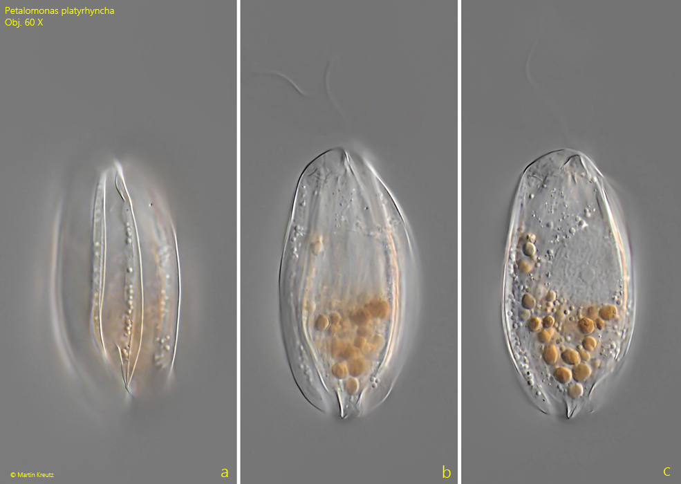
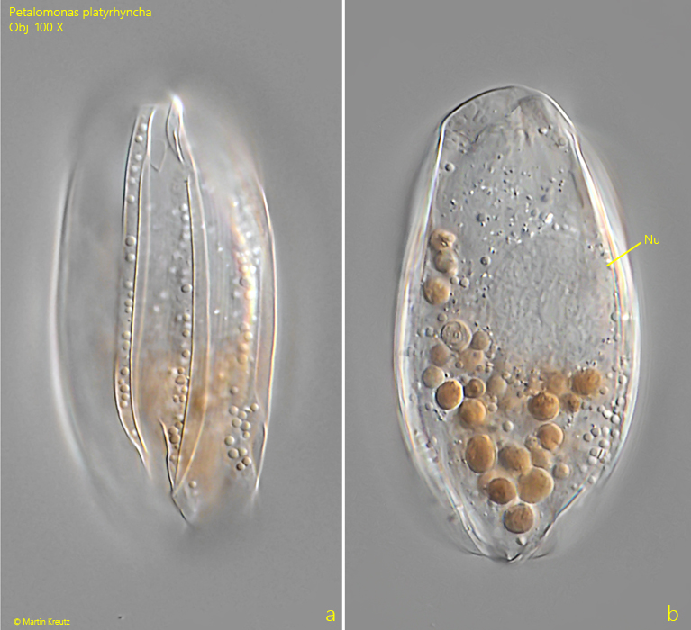
Petalomonas platyrhyncha is a very common species in the Simmelried. Almost every sample contains specimens. The population in Simmelried seems to be larger than the one described by Skuja from Swedish Lapland. He gives a maximum length of 48 µm. However, I could also find specimens with more than 70 µm length. Thus, Petalomonas platyrhyncha belongs to one of the largest Petalomonas species. The cells are mostly filled with colorless or orange, highly refractive spheres. The nucleus is large and centrally located. The species has a round, snout-shaped apex, which reliably distinguishes it from the similar species Petalomonas praegnans, which is symmetrically pointed at the anterior end.
Fig. 1 a-c: Petalomonas platyrhyncha. L = 59 µm. Three focal planes of a freely swimming specimen from dorsal. Obj. 60 X.
Fig. 2 a-b: Petalomonas platyrhyncha. L = 59 µm. The same specimen as shown in fig. 1-a-c flightly squashed. Nu = nucleus. Obj. 100 X.
Fig. 3 a-c: Petalomonas platyrhyncha. L = 71 µm. A second freely swimming specimen from dorsal (a) and in lateral view from left (b, c). AP = apex, F = flagellum, K1–K3 = dorsal keels. Obj. 100 X.
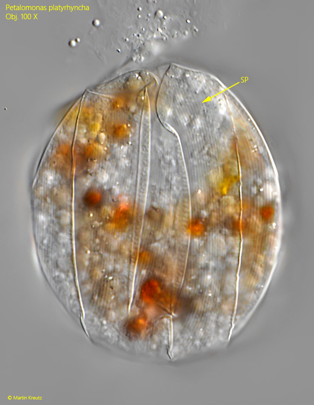
Fig. 4: Petalomonas platyrhyncha. A squashed specimen from dorsal with focal plane on the delicate striation of the pellicle (SP). Obj. 100 X.
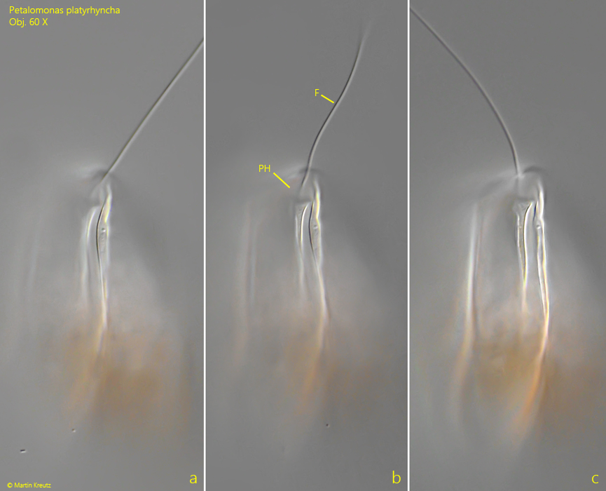
Fig. 5: Petalomonas platyrhyncha. The pharynx (PH) and the flagellum (F) in detail. Obj. 60 X.
Fig. 6: Petalomonas platyrhyncha. L = 52 µm. Lateral view of a specimen during cell division. Obj. 40 X.