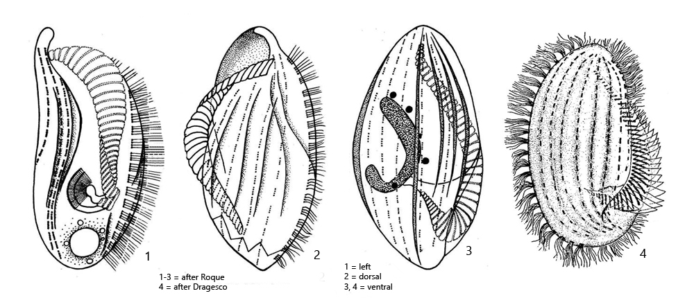
4–5 longitudinal ridges on ventral and dorsal side
length 70–140 µm
prominent adoral zone of membranelles running from anterior end over the left side to the mouth opening on ventral side
mouth opening in posterior quarter
macronucleus horseshoe-shaped
5–9 micronuclei adjacent to macronucleus
contractile vacuole terminal
Phacodinium metchnikoffi
I find Phacodinium metchnikoffi frequently and regularly in moss samples. This ciliate seems to be particularly common in moss growing on trees. Kahl has found Phacodinium metchnikoffi in moss growing on roofs.
Phacodinium metchnikoffi is very conspicuous due to its long adoral zone on the left side of the body and the longitudinal ribs on both sides of the body. This makes it conspicuous even at low magnifications. The mouth opening is in the posterior quarter. The specimens of my population with a length of 130–140 µm were at the upper end of the range given by Kahl and Penard.
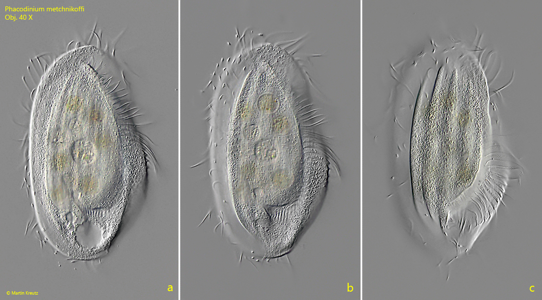
Fig. 1 a-c:Phacodinium metchnikoffi. L = 141 µm. Different focal planes of a freely swimming specimen from ventral. Obj. 40 X.
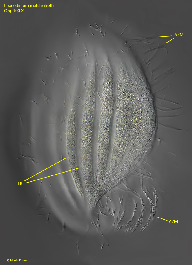
Fig. 2:Phacodinium metchnikoffi. L = 130 µm. A slightly squashed specimen from ventral with focal plane on the longitudinal ridges (LR). Note the long adoral zone of membranelles (AZM) running over the left side to the ventral side in the posterior quarter. Obj. 100 X.
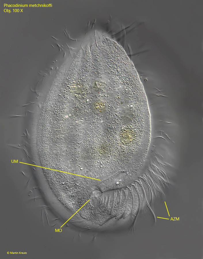
Fig. 3:Phacodinium metchnikoffi. L = 130 µm. The same specimen as shown in fig. 2 with focal plane on the mouth opening (MO). Note the narrow groove that leads diagonally upwards at the edge of which the undulating membrane (UM) lies. AZM = adoral zone of membranelles. Obj. 100 X.
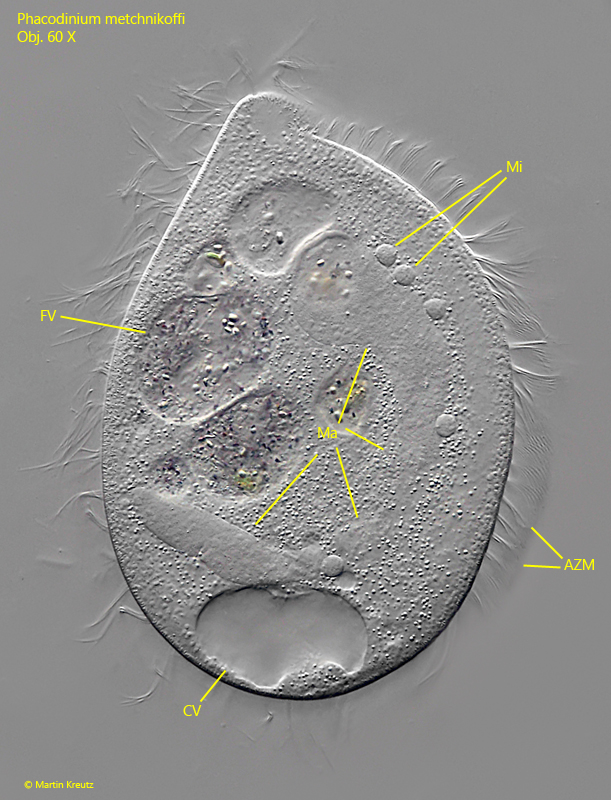
Fig. 4:Phacodinium metchnikoffi. The horseshoe-shaped macronucleus (Ma) and the adjacent micronuclei (Mi) in a strongly squashed specimen. Obj. 100 X.