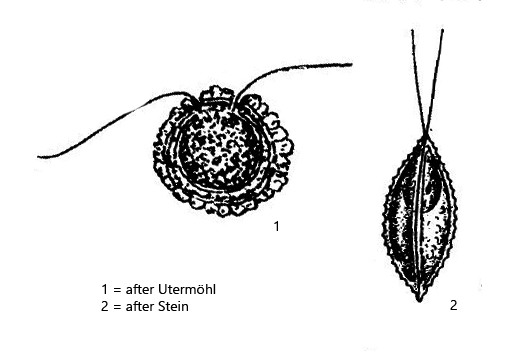
shell lenticular in lateral view with an outer rim
shell is composed of two halves
shell thick, calcified, two or more layers, colorless, brownish or yellowish
two flagella
one eyespot in mid-cell
one chloroplast with one pyrenoid
contractile in hyaline anterior end
Phacotus lenticularis
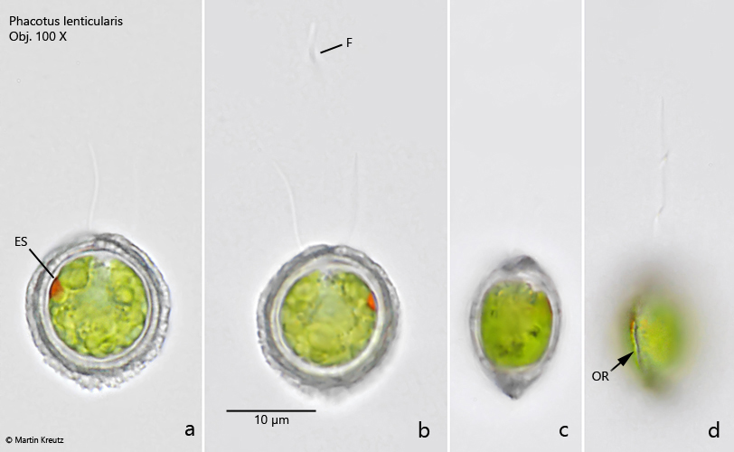
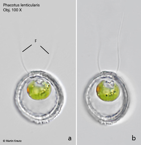
Phacotus lenticularis is a volvococcal alga with a planktonic lifestyle. I found it in October 2022 in the plankton of Mühlhalden pond. With about 15 µm diameter of the calcified shell, the alga is very small, but often occurs in masses. Often the shells are distinctly brown or even opaque. In my population, however, many bright and transparent specimens were present. Phacotus lenticularis is best observed under brightfield illumination because the mineral calcite inclusions of the shell shine very brightly in DIC. The shell consists of several layers. The outermost layer is large and has an almost crystalline character. The two flagella are not easy to detect in brightfield illumination. It is not clear from the literature whether both flagella arise from one opening, or from two separate ones. According to my observations there are two separate openings. This can also be seen from the fact that the flagella are quite far apart (s. fig. 2 a-b). The lenticular shell consists of two halves. A thin rim runs along the contact surface of the shell halves (s. fig. 1d). Only during reproduction the two halves of the shell move apart to release the daughter cells.
Fig. 1 a-d:Phacotus lenticularis. D = 15 µm. Frontal view (a, b) and lateral view (c, d) of a freely swimming specimen. Note the outer rim (OR, fig. 1d) of the shell where both halves of the shell are joined. ES = eye spot, F = flagellum. Obj. 100 X.
Fig. 2 a-b:Phacotus lenticularis. D = 16 µm. Frontal view of a second, freely swimming specimen with a smaller protoplast. The two flagella (F) arise separately, each from the side of an apical thickening of the shell. Obj. 100 X.
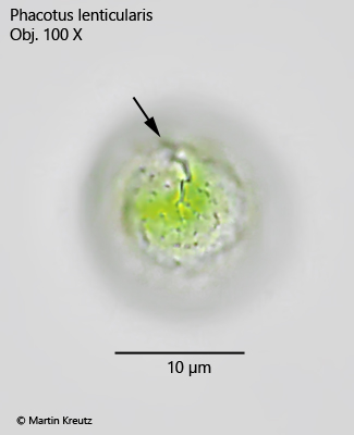
Fig. 3:Phacotus lenticularis. Focal plane on the surface of the calcified shell. Under increasing pressure of the coverslip the shell starts to crack (arrow). Obj. 100 X.