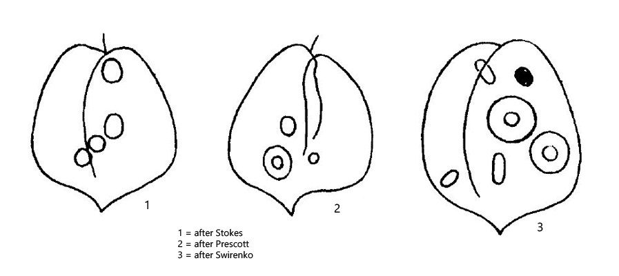
Phacus acuminatus can is a very common species and I find it in different sites. The species can be recognized by its broad oval shape and the very short, V-shaped spine (s. fig. 1b). Usually 1–2 large, disc-shaped paramylon bodies are present in the cell, sometimes with a central hole or even ring-shaped. The specimens in my population were mostly 30 µm long. Because of the small size the species is often overlooked.
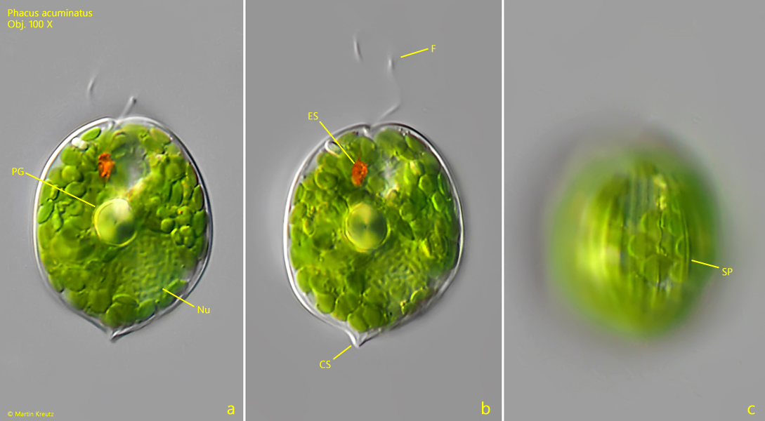
Fig. 1 a-c:Phaus acuminatus. L = 30 µm. Different focal planes of a slightly squashed specimen. CS = caudal spine, ES = eyespot, F = flagellum, Nu = nucleus, PG = prominent paramylon grain, SP = striation of pellicle. Obj. 100 X.
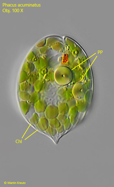
Fig. 2:Phaus acuminatus. L = 32 µm. A second, strongly squashed specimen with two prominent paramylon bodies (PP). Chl = disc-shaped chloroplasts. Obj. 100 X.