cell pear-shaped or ovoid, dorso-ventrally flattened
dorsal keel with length of cell
length 31–50 µm, width 15–27 µm
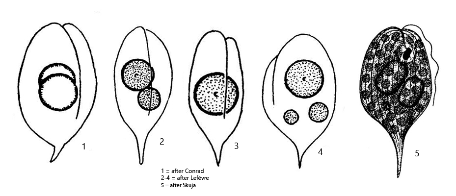
1–2 prominent paramylon bodies, oval or circular
chloroplasts disc-shaped
posterior end tapering continuously into a short caudal spine
caudal spine, 5–11 µm long, straight or slightly curved
one flagellum, about body length
pellicle longitudinally striated
eyespot present
Phacus caudatus
I regularly find Phacus caudatus, but usually only isolated cells. I recognize the species mainly by the short caudal spine, which is formed by a continuous tapering of the posterior end and which is mostly straight or only slightly bent. Also, this species has a dorsal keel that is very pronounced and runs along the entire cell (s. figs. 1 b and 2 d). The cell shape is quite variable. I have found slender, almost parallel-sided specimens. In my population the specimens were never longer than 45 µm.
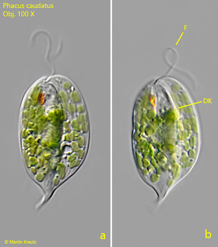
Fig. 1 a-b:Phacus caudatus. L = 37 µm. Two focal planes of a freely swimming specimen from dorsal. Note the dorsal keel (DK) running along the whole cell. F = flagellum. Obj. 100 X.
Fig. 2 a-d:Phacus caudatus. L = 43 µm. Different focal planes of a second specimen from ventral. In figs. c and d is the focal plane on the dorsal side. Chl = disc-shaped chloroplasts, CV = contractile vacuole, DK = dorsal keel, ES = eyespot, F = flagellum, Nu = nucleus. Obj. 100 X.