longitudinally obvoid in outline, slightly asymmetric
anterior “lips” overlapping
narrowing abruptly in a long spine, spine often bent
pellicle longitudinally striated
red eyespot prominent
chloroplasts disc shaped
paramylon in small discs or 1–2 large discs
flagellum shorter than cell
pyrenoids absent
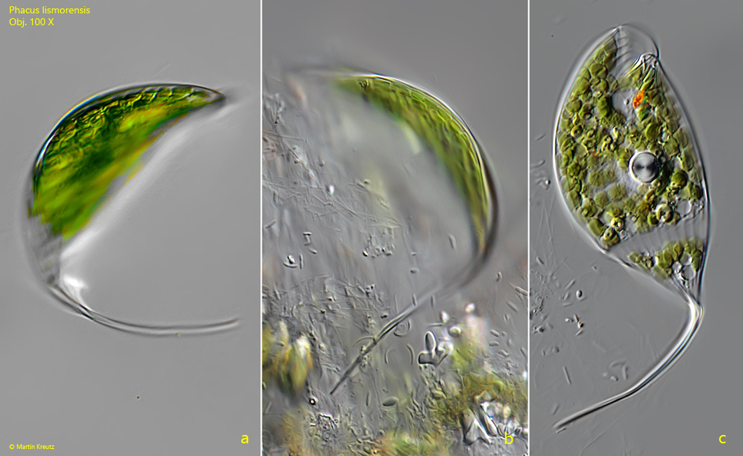
Phacus lismorensis
I found the first specimens of Phacus lismorensis in 1994 in Simmelried and later also in Ulmisried. In both locations the species is very common. The cells are typically asymmetrically shaped with a rapidly tapering spine. The cells can appear very curved and bent (s. fig. 3 a-c). The shape of the paramylon grains did not seem constant to me. I found specimens its with larger, disc-shaped paramylon as well as those in which it was present in many small discs.
Fig. 1 a-c:Phacus lismorensis. L = 154 µm. Three focal planes of an unsquashed specimen. ES = eyespot, Nu = nucleus. Obj. 100 X.
Fig. 2 a-c:Phacus lismorensis. L = 141 µm. A second, slightly squashed specimen. Note the large paramylon grain (LGP) filling almost the whole body and the small paramylon grains (SPG). Nu = nucleus. Obj. 60 X.
Fig. 3 a-c:Phacus lismorensis. L = 105 µm. a, b) a strongly bent specimen. c) the same specimen strongly squashed. Obj. 100 X.
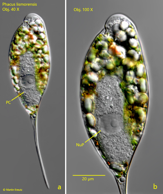
In many samples I found infestation of Phacus lismorensis by a parasitic fungus. In most cases it was only one very large parasitic cell, which was intracellular (s. fig. 4 a-b). Sometimes there were even two parasitic cells. Reddish-brown metabolites were deposited in these cells and after prolonged infestation, the paramylon granules were completely degraded to feed the parasite. Unfortunately, I could not observe the complete life cycle of the parasitic fungus.
Fig. 4 a-b:Phacus lismorensis. L = 88 µm. A specimen that has been infested by a parasitic fungus. PC = parasitic cell, NuP = nucleus of the parasitic cell. Obj. 100 X.