body cylindrical, anterior third ventrally bent, posterior end rounded
length 25–35 µm
oral aperture rounded, surrounded by cilia
pharyngeal basket present
pellicle rigid, longitudinally ribbed
somatic cilia reduced to a ventral field of cilia located at posterior third
macronucleus spherical in centre or anterior half
one caudal cilium
Pithothorax rotundus
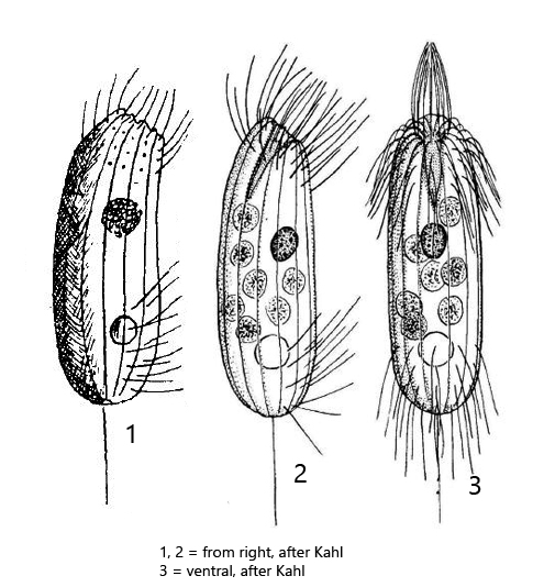
I found Pithothorax rotundus twice in short succession in November and December 2022 in floating, decaying plant masses in the Simmelried. This ciliate is quite small with a length of 25–35 µm (according to Kahl) and also occurs only sporadically. Possibly I missed it in earlier samples, although the body shape with the ventrally bent anterior end is very characteristic. The cytopharynx is supported by very fine, rod-like trichites. The ciliature is very reduced. There is a ring of cilia around the mouth opening and a small field of cilia is found on the ventral side in the posterior third (s. figs. 1b, 1c, 2b and 3a). Beyond that, only a single caudal cilium is found, which is hard to see (s. figs. 3b and 3c). Kahl drew the position of the spherical macronucleus in the middle of the cell. In my specimens it was positioned at the posterior end (s. fig. 2a). The contractile vacuole is found in the posterior third of the body as described by Kahl (s. fig. 2b). My specimens with 36–38 µm length were at the upper limit of the body length given by Kahl, but still within the variability.
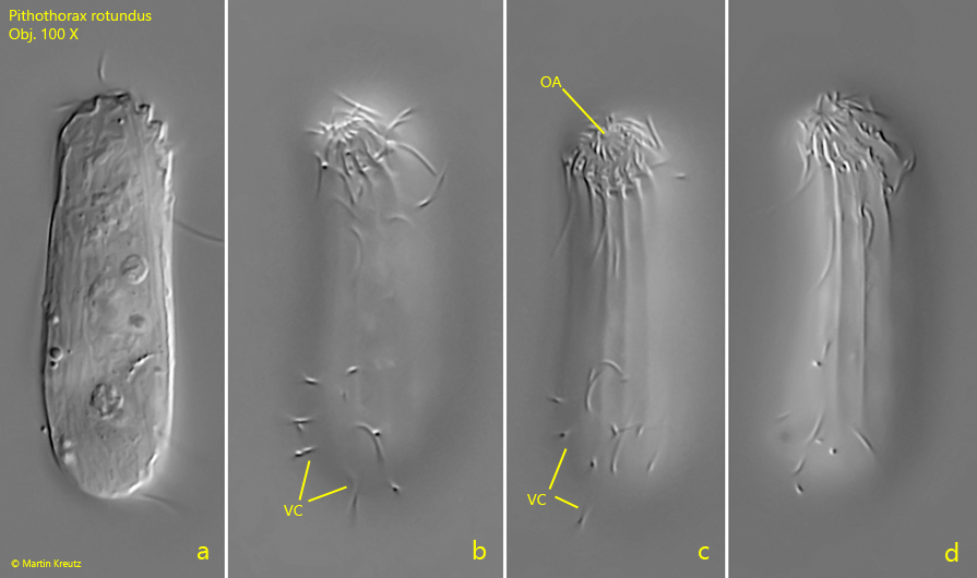
Fig. 1 a-d: Pithothorax rotundus. L = 38 µm. Four focal planes of the ventral side of a freely swimming specimen. Note the cilia surrounding the oral aperture (OA) and the field of ventral cilia (VC) in the posterior third. Obj. 100 X.
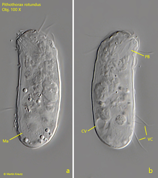
Fig. 2 a-b: Pithothorax rotundus. L = 38 µm. The same specimen as shown in fig. 1 a-d from the left side (a) and from the right side. In the center of the macronucleus (Ma) a nucleolus is visible. The contractile vacuole (CV) is located dorsally in the posterior quarter of the cell. PB = pharyngeal basket, VC = field of ventral cilia. Obj. 100 X.
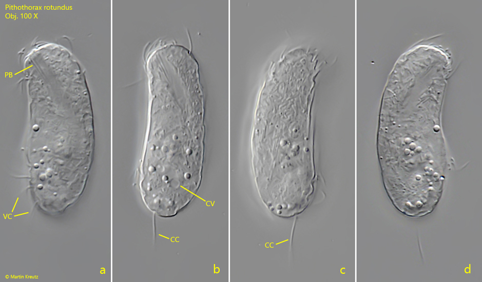
Fig. 3 a-d: Pithothorax rotundus. L = 36 µm. A second freely swimming specimen from left (a, b) and from right (c, d). CC = caudal cilium, CV = contractile vacuole, PB = pharyngeal basket, VC = field of ventral cilia. Obj. 100 X.