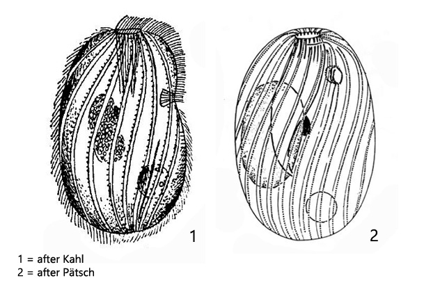
lateral invgination of unknown function in anterior third
macronucleus oval
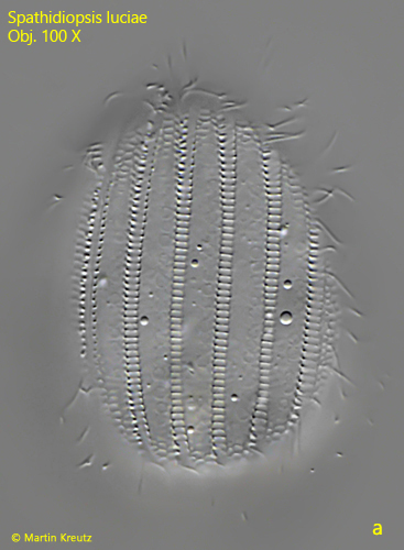
pellicle with conspicuously spiral ridges
contractile vacuole located ventral and subterminal
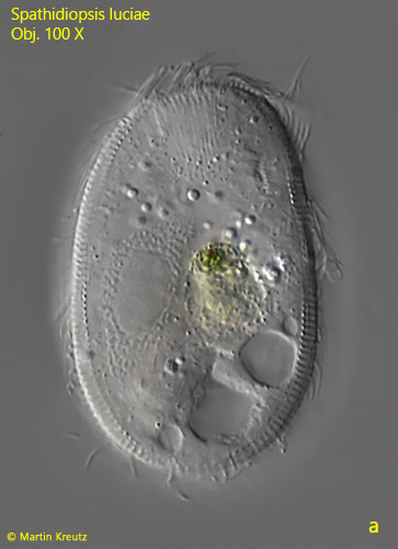
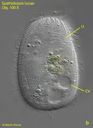
Spathidiopsis luciae
Spathidiopsis luciae is described as a very common ciliate. However, since 1992 I have only been able to find one specimen in Ulmisried in the year 2022. There I found the specimen in a sample from an accumulation of leaves, which were jammed in front of a weir overflow. Spathidiopsis luciae is quite small at about 50 µm, but it is immediately recognizable by fielded and ribbed stripes arranged parallel to the kineties. The purpose of this fielding is unknown. In addition, it also possesses a laterally located invagination (LI). The function of the invagination is also unknown and it is not associated with the mouth opening (MO):
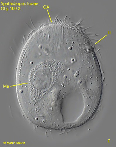
Fig. 1 a-c:Spathidiopsis luciae. L = 47 µm. a,b) slightly squashed specimen. c) strong squashed specimen. CV = contractile vacuole, Ma = macronucleus, LI = lateral invagination, OA = oral apparatus. Obj. 100 X.
Fig. 2 a-b:Spathidiopsis luciae. L = 47 µm. The fielded and ribbed stripes of the pellicle in two focal planes. Obj. 100 X.