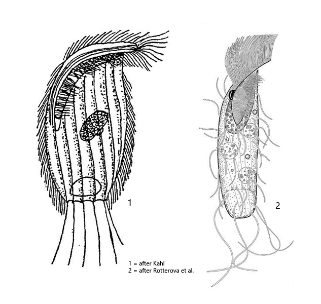
body broadly oval, almost recangular, dorso-ventrally flattened
apical dome flattened and bent ventrally
length 60–100 µm
adoral zone limited to ventral side, short, with only few membranelles
perizonal stripe of 5 ciliary rows, shorter than adoral zone
macronucleus oval with adjacent micronucleus
rows of somatic cilia are wide spaced und run obliquely counterclockwise
large contractile vacuole terminal
truncated posterior end with long caudal cilia
Planometopus contractus
Planometopus contractus was first described by Penard in 1922 as Metopus contractus. This name was adopted by Kahl. In 2018, Rottovera et al. redescribed the species and transferred it to the newly established genus Planometopus based on morphological characters and the results of 18S rRNA gene analysis.
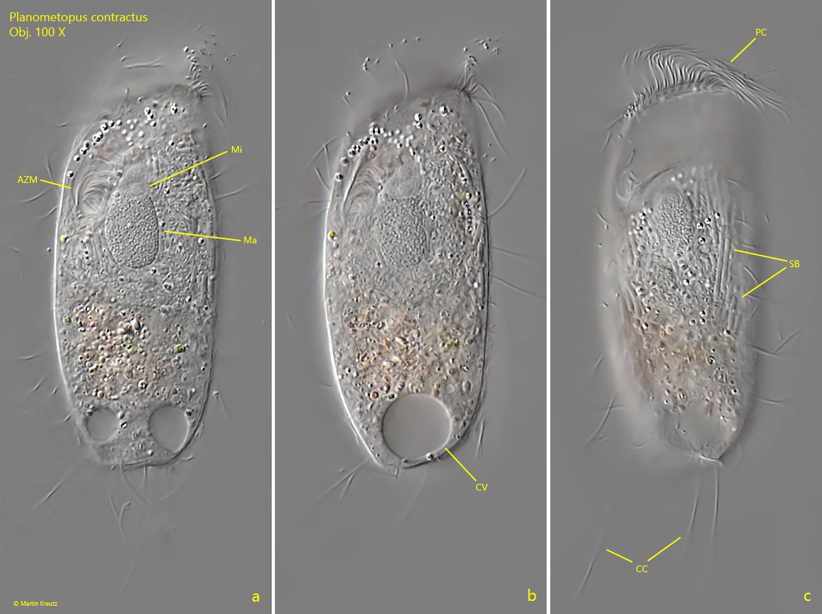
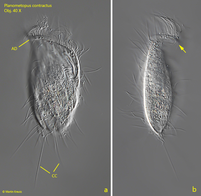
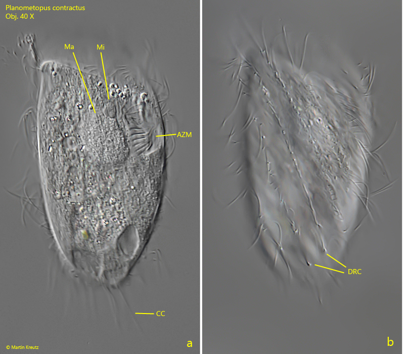
So far I could detect Planometopus contractus exclusively in the Simmelried where the species occurs sporadically. The shape of Planometopus contractus is very variable, which is also mentioned by Kahl and Rottovera et al. I found stocky, almost rectangular specimens (s. fig. 3 a-b) and also more elongate, oval specimens (s. fig. 1 a-c). However, all forms are distinctly dorso-ventrally flattened (s. fig. 2 b) and the apical dome is flat and distinctly curved ventrally (s. fig. 2 b). Overall, the ciliate appears very rigid and not flexible. What is also striking compared to other metopid ciliates are the widely spaced rows of somatic cilia, which are running obliquely and in a counterclockwise manner. This can be seen especially clearly on the dorsal side (s. fig. 3 b). In my population, I could observe very large symbiotic bacteria in the cytoplasm, which accumulated around the macronucleus (s. fig. 1 c). Since these symbiotic bacteria are not mentioned by Kahl or Rottovera et al. it is unclear whether this is a constant feature. The long caudal cilia appeared spread in a fan-shaped way in some specimens, whereas they were soft and flexible in others.
Fig. 1 a-c:Planometopus contractus. L = 72 µm. A freely swimming specimen from ventral. AZM = adoral zone of membranelles, CC = caudal cilia, CV = contractile vacuole, Ma = macronucleus, Mi = micronucleus, PC = perizonal cilia, SB = symbiotic bacteria. Obj. 100 X.
Fig. 2 a-b:Planometopus contractus. L = 94 µm. A freely swimming specimen from dorsal (a) and from left (b). Note the ventrally bent apical dome (arrow). AD = apical dome, CC = caudal cilia. Obj. 40 X.
Fig. 3 a-b:Planometopus contractus. L = 75 µm. A freely swimming specimen from dorsal. AZM = adoral zone of membranelles, CC = caudal cilia, DRC = dorsal rows of somatic cilia, Ma = macronucleus, Mi = micronucleus. Obj. 100 X.