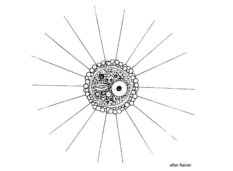
nucleus located eccentrically, with central nucleolus
no contractile vacuole visible
pseudopodia fine and short
Pompholyxophrys punicea
I find Pompholyxophrys punicea regularly, but never frequently in some of my sites. Mostly I find specimens at the bottom of old samples with little plant material. The cells can be recognized by their orange coloration even at low magnification. However, the beautiful pearly scales surrounding the cell body can only be seen at high magnifications. Often the spherules are arranged in multiple, concentric layers, with the smallest spherules forming the innermost layer. The spherical scales are formed by the cell itself and consist of amorphous silicium.
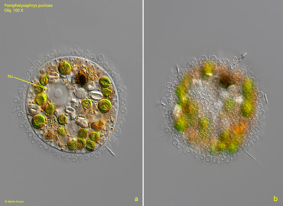
Fig. 1 a-b:Pompholyxophrys punicea. D = 47 µm (without scales). A squashed specimen with a pale orange color. Note the layers of transparent spherules (scales) covering the cell. Nu = nucleus, PP = pseudopodia. Obj. 100 X.
Fig. 2 a-b:Pompholyxophrys punicea. D = 50 µm (without scales). A second, slightly squashed specimen with a large number of ingested algae. Nu = nucleus. Obj. 100 X.
Fig. 3 a-b:Pompholyxophrys punicea. D = 35 µm (without scales). A third, slightly squashed specimen with extended pseudopodia. Obj. 100 X.